Anatomy & Physiology 2
Subtopic:
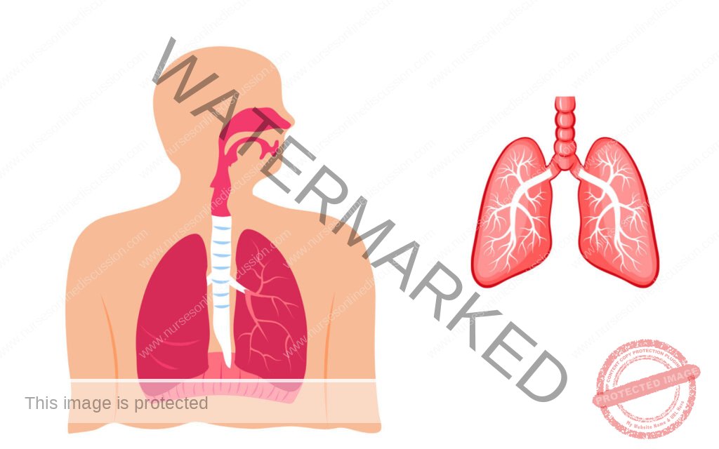
The Respiratory System
The Respiratory System
Objectives;
- At the end of the chapter, the student should be able to:
- Describe the purpose of the respiratory system
- Differentiate between external and internal respiration
- Name all of the structures of the respiratory system
- Explain how food and foreign materials are kept out of the respiratory tract
- Explain the mechanism for the pulmonary ventilation
- List and define five breathing volumes;
Notes
General Function
- A primary requirement for all body cell activities and growth is oxygen, which is needed to obtain energy from food. The fundamental purpose of the respiratory system is to supply oxygen to the individual tissue cells and to remove their gaseous waste product, carbon dioxide. Breathing, or ventilation, refers to the inhalation and exhalation of air.
- Air is a mixture of oxygen, nitrogen, carbon dioxide and other gases; the pressure of these gases varies, depending on the elevation above sea level. The first, called external expiration, takes place only in the lungs, where oxygen from the outside air enters the blood and carbondioxide leaves the blood to be breathed into the outside air.
- In the second, called internal respiration, gas exchanges take place between the blood and the body cells, with oxygen leaving the blood and entering the cells at the same time that carbon dioxide leaves the cells and enters the blood.
- The respiratory system is an intricate arrangement of spaces and passageways that conduct air into the lungs. These spaces include the nasal cavities; the pharynx, which is common to the digestive and respiratory systems; the voice box, or larynx; the windpipe, or trachea; and the lungs themselves, with their conducting tubes and air sacs. The entire system might be thought of as a pathway for air between the atmosphere and the blood.
Structure and Function of Respiratory Pathways;
The Nasal Cavities;
Air makes its initial entrance into the body through the openings in the nose called the nostrils. Immediately inside the nostrils, located between the roof of the mouth and the cranium, are the two spaces known as the nasal cavities.
NB, Draw the diagram of each organ described;
These two spaces are separated from each other by a partition, the nasal septum. The septum and the walls of the nasal cavities are constructed of bone covered with mucous membrane. From the lateral (side) walls of each nasal cavity are three projections called the conchae. The conchae greatly increase the surface over winch air must travel on its way through the nasal cavities.
The lining of the nasal cavities is a mucous membrane, which contains many blood vessels that bring heat and moisture to it. The cells of this membrane secrete a large amount of fluid.
It is better to breathe through the nose than through the mouth because of changes produced in the air as it comes in contact with the lining of the nose:
- Foreign bodies, such as dust particles and pathogens, are filtered out by the hairs of the nostrils or caught in the surface mucus.
- Air is warned by the blood in the vascular membrane.
- Air is moistened by the liquid secretion
The sinuses are small cavities lined with mucous membrane in the bones of the skull. The sinuses communicate with the nasal cavities, and they are highly susceptible to infection.
The Pharynx
The muscular pharynx (throat) carries air into the respiratory tract and foods and liquids into the digestive system. The upper portion located immediately behind the nasal cavity is called the nasopharynx, the middle section located behind the mouth is called the oropharynx, and the lowest portion is called the laryngeal pharynx. This last section opens into the larynx toward the front and into the oesophagus toward the back.
The Larynx
- The larynx (voice box) is located between the pharynx and the trachea. It has a framework of cartilage that protrudes in the front of the neck and sometimes is referred to as the Adam’s apple. The larynx is considerably larger in the male than in the female; hence, the Adam’s apple is much more prominent in the male. At the upper end of the larynx are the vocal cords, which serve in the production of speech. They are set into vibration by the flow of air from the lungs.
- A difference in the size of the larynx is what accounts for the difference between the male and female voices; because a man’s larynx is larger than a woman’s, his voice is lower in pitch. The nasal cavities, the sinuses, and the pharynx all serve as resonating chambers for speech, just as the cabinet does for a stereo speaker.
- The space between these two vocal cords is called the glottis, and the little leaf-shaped cartilage that covers the larynx during swallowing is called the epiglottis. The epiglottis helps keep food out of the remainder of the respiratory tract. As the larynx moves upward and forward during swallowing, the epiglottis moves downward, covering the opening into the larynx. You can feel the larynx move upward toward the epiglottis during this process by placing the flat ends of your fingers on your larynx as you swallow. Draw the
structure of the vocal cords:
The larynx is lined with ciliated mucous membrane. The cilia trap dust and other particles, moving them upward to the pharynx to be expelled by coughing, sneezing, or blowing the nose.
The Trachea (Windpipe) diagram
The trachea is a tube that extends from the lower edge of the larynx to the upper part of the chest above the heart. It has a framework of cartilages to keep it open. These cartilages, shaped somewhat like a tiny horseshoe or the letter C, are found along the entire length of the trachea. All the open sections of these cartilages are at the back so that the esophagus can bulge into this section during swallowing. The purpose of the trachea is to conduct air between the larynx and the lungs.
The Bronchi and Bronchioles
- The trachea divides into two bronchi which enter the lungs.
- The right bronchus is considerably larger in diameter than the left and extends downward in a more vertical direction.
- Therefore, if a foreign body is inhaled, it is likely to enter the right lung. Each bronchus enters the lung at a notch or depression called the hilus or hilum. The blood vessels and nerves also connect with the lung in this region.
The Lungs
Draw the structure of the lungs
- The lungs are the organs in which external respiration takes place through the extremely thin and delicate lung tissues.
- The two lungs, set side by side in the thoracic cavity, are constructed in the following manner:
- Each bronchus enters the lung at the hilus and immediately subdivides. Because the subdivision of the bronchi resembles the branches of a tree, they have been given the common name bronchial tree. The bronchi subdivide again and again, forming progressively smaller divisions, the smallest of which are called bronchioles. The bronchi contain small bits of cartilage, which give firmness to the walls and serve to hold the passageways open so that air can pass in and out easily.
- However, as the bronchi become smaller, the cartilage decreases in amount. In the bronchioles there is no cartilage at all; what remains is mostly smoothly muscle, which is under the control of the autonomic nervous system.
- At the end of each of the smallest subdivisions of the bronchial tree, called terminal bronchioles, is a cluster of air sacs, resembling a bunch of grapes. These sacs are known as alveoli. Each alveolus is a single-cell layer of squamous (flat) epithelium.
- This very thin wall provides easy passage for the gases entering and leaving the blood as it circulates through millions of tiny capillaries of the alveoli. Certain cells in the alveolar wall produce surfactant, a substance that prevents the alveoli from collapsing by reducing the surface tension (“pull”) of the fluids that line them. There are millions of alveoli in the human lung. Because of the many air spaces, the lung is light in weight; normally a piece of lung tissue dropped into a glass of water will float.
As mentioned the pulmonary circuit brings blood to and from the lungs. In the lungs blood passes through the capillaries around the alveoli, where the gas exchange takes place.
The Lung Cavities
- The lungs occupy a considerable portion of the thorax cavity, which is separated from the abdominal cavity by the muscular partition known as the diaphragm. Each lung is enveloped in a double sac of serous membrane called the pleura. The portion of the pleura that is attached to the chest wall is called parietal pleura, while the portion that is reflected onto the surface of the lung is called visceral pleura.
- The pleural cavity around the lungs is an air-tight space with a partial vacuum, which causes the pressure in this space to be less than atmospheric pressure. Because the pressure inside the lungs is higher than that in the surrounding pleural cavity, the lungs tend to remain inflated. The entire thoracic cavity is flexible, capable of expanding and contracting along with the lungs. The region between the lungs, the mediastinum, contains the heart, great blood vessels, esophagus, trachea, and lymph nodes.
Physiology of Respiration (mechanisms of respiration)
Pulmonary Ventilation
Ventilation is the movement of air into and out of the lungs, as in breathing. There are two phases of ventilation:
- Inhalation is the drawing of air into the lungs.
- Exhalation is the expulsion of air from the lungs.
In inhalation, the active phase of breathing, the respiratory muscles contract to enlarge the thoracic cavity. The diaphragm is a strong dome-shaped muscle attached around the base of the rib cage. The contraction and relaxation of the diaphragm cause a piston-like downward motion that result in an increase in the vertical dimension of the chest. The rib cage is also moved upward and outward by contraction of the external intercostals muscles and, during exertion, by contraction of other muscles of the neck and chest. During quiet breathing, the movement of the diaphragm accounts for most of the increase in thoracic volume.
As the thoracic cavity increases in size, gas pressure within the cavity decreases. When the pressure drops to slightly below atmospheric pressure, air is drawn into the lungs.
In exhalation, the passive phase of breathing, the muscles of respiration relax, allowing the ribs and diaphragm to return to their original positions. The tissues of the lung are elastic and recoil during exhalation. During forced exhalation, the internal intercostals muscles and the muscles of the abdominal wall contracts, pulling the bottom of the rib cage in and down. The abdominal viscera are also pushed upward against the diaphragm.
Air Movement
Air enters the respiratory passages and flows through the ever-dividing tubes of the bronchial tree. As the air traverses this passage, it moves more and more slowly through the great number of bronchial tubes until there is virtually no forward flow as it reaches the alveoli. Here the air moves by diffusion, which soon equalizes any differences in the amounts of gases present. Each breath causes relatively little change in the gas composition of the alveoli, but normal continuous breathing ensures the presence of adequate oxygen and the removal of carbon dioxide.
External respiration:
Draw the diagram of external respiration
This is the exchange of gases between the alveoli and the blood in the alveolar capillaries, across the respiratory membrane. Each alveolar wall is surrounded by a net work of tiny capillaries (the walls of which are only one cell thick). Venous blood arriving at the lungs has travelled from all the tissues of the body, and contains high levels of CO2 and low levels of O2. Carbon dioxide diffuses from the venous blood down its concentration gradient into the alveoli until equilibrium with alveolar air is reached. By the same way O2 diffuses from the alveoli into the blood.
Internal respiration
This is exchange of gases by diffusion between the blood in the capillaries and the body cells (draw the diagram)
Breathing Volumes:
Tidal volume – The amount of air moved into or out of the lungs in quiet, relaxed breathing
500 cc
Inspiratory reserve volume– this is the extra volume of air that can be inhaled in the lungs during maximum inspiration, i.e. over and above normal TV.
Inspiratory capacity- this is the amount of air that can be inspired with maximum effort. It consists of the tidal volume (500mls) plus the inspiratory reserve volume.
Vital capacity– The volume of air that can be expelled from the lungs by maximum exhalation following maximum inhalation 4800 cc
Residual volume– The volume of air that remains in the lungs after maximum exhalation 1200 cc
Total lung capacity– The total volume of air that can be contained in the lungs after maximum inhalation 6000 cc
Functional residual capacity – The amount of air remaining in the lungs after normal exhalation
2400 cc
Regulation of respiration
- Regulation of respiration is a complex process that must keep pace with moment-to-moment changes in cellular oxygen requirements and carbon dioxide production. Regulation depends primarily on the respiratory control centers located in the medulla and pons of the brain stem. Nerve impulses from the medulla are modified by the centers in the pons.
- Respiration is regulated so that the levels of oxygen, corbondioxide, and acid are kept within certain limits. The control centers regulate the rate, depth, and rhythm of respiration.
- From the respiratory center in the medulla, motor nerve fibers extend into the spinal cord. From the cervical (neck) part of the cord, these nerve fibers continue through the phrenic nerve to the diaphragm.
- The diaphragm and the other muscles of respiration are voluntary in the sense that they can be regulated by messages from the higher brain centers, notably the cortex. It is possible for a person to deliberately breathe more rapidly or more slowly or to hold his breath and not breathe at all for a time.
- Usually we breathe without thinking about it, while the respiratory centers in the medulla and pons do the controlling.Of vital importance in the control of respiration are the chemoreceptors. These receptors are found in structures called the carotid and aortic bodies, as well as outside the medulla of the brain stem. The carotid bodies are located near the bifurcation of the common carotid arteries, while the aortic bodies are located in the aortic arch.
- These bodies contain many small blood vessels and sensory neurons, which are sensitive to decreases in oxygen supply as well as to increases in carbon dioxide and acidity (H+). Impulses are sent to the brain from the receptors in the carotid and aortic bodies. The receptor cells outside the medulla are affected by the concentration of hydrogen ion in cerebrospinal fluid (CSF) as determined by the concentrations of carbon dioxide in the blood.
We are a supportive platform dedicated to empowering student nurses and midwives through quality educational resources, career guidance, and a vibrant community. Join us to connect, learn, and grow in your healthcare journey
Quick Links
Our Courses
Legal / Policies
Get in Touch
(+256) 790 036 252
(+256) 748 324 644
Info@nursesonlinediscussion.com
Kampala ,Uganda
© 2026 Nurses online discussion. All Rights Reserved