Anatomy & Physiology 2
Subtopic:
Organs of Special Senses
Organs of Special Senses
Sensation is a conscious or sub conscious awareness of external or internal stimuli. The special senses like the somatic senses allow us to detect specific changes in our environment. However, the special senses like smell, taste, vision, hearing and equilibrium provide richer sensory experiences. They also have receptor organs that are structurally more complex. These senses and their receptors include;
Olfactory Sensation
Both smell and taste are chemical senses; that is the sensations arise from the interaction of molecules with smell or taste receptors. Among all sensations, only smell and taste project both higher cortical areas and to the limbic system. This is probably the reason that curtain orders and tests can evoke strong emotional responses as a flood of memories.
Anatomy of olfactory receptors
About 12 million receptors for the olfactory sense lie in the nasal epithelium in the superior portion of the nasal cavity. The total area of the olfactory epithelium is 5cm. it occupies the superior portion of the nasal cavity extending along the superior nasal concha and upper part of the middle nasal concha. The olfactory epithelium consists of three kinds of cells i.e. olfactory receptors, supporting cells and basal cells.
SPECIAL SENSES
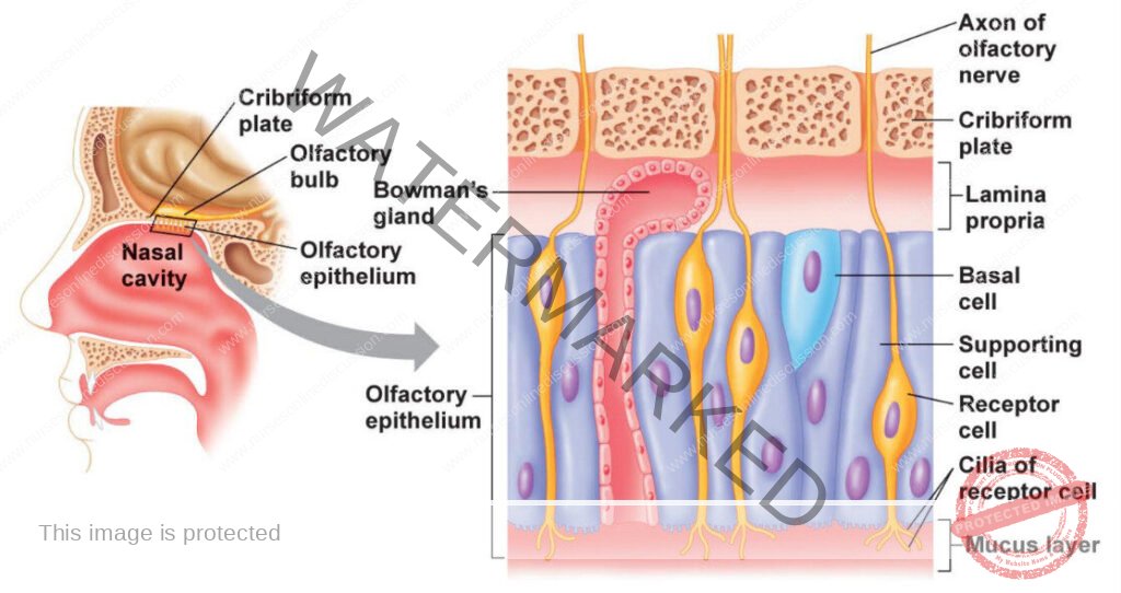
Olfactory receptors are bipolar neurons whose distal end is a Knob shaped dendrite. Several cilia, called olfactory hairs project from the dendrite. The cilia act as sites for olfactory transduction. Olfactory receptors respond to chemical stimulation of an adorant molecule by providing a generator potential thus initiating the response. For the proximal part of each olfactory receptor, a single axon projects to the olfactory bulb. Supporting cells are columnar epithelial cells of the mucus membrane lining the nose. Basal cells lie between the bases of supporting cells. They are stem cells that continually produce new olfactory receptors which live for only one month and they are replaced.
Within the connective tissue that support the olfactory epithelium are olfactory (bowman’s glands). They produce mucus that is carried to the surface of the epithelium by ducts. This secretion moistens the surface of the olfactory epithelium and dissolves adorant gases. Both supporting cells of the nasal epithelium and olfactory glands are innervated by branches of the facial nerve V.
TASTE SENSATION
The taste buds, the sensory receptors for taste are located primarily in the oral cavity. Of our 10000 or so taste buds, most are on the tongue. A few are scattered on the soft palate, inner surface of the cheeks, pharynx and the epiglottis of the larynx.
ANATOMY OF THE TASTE BUDS
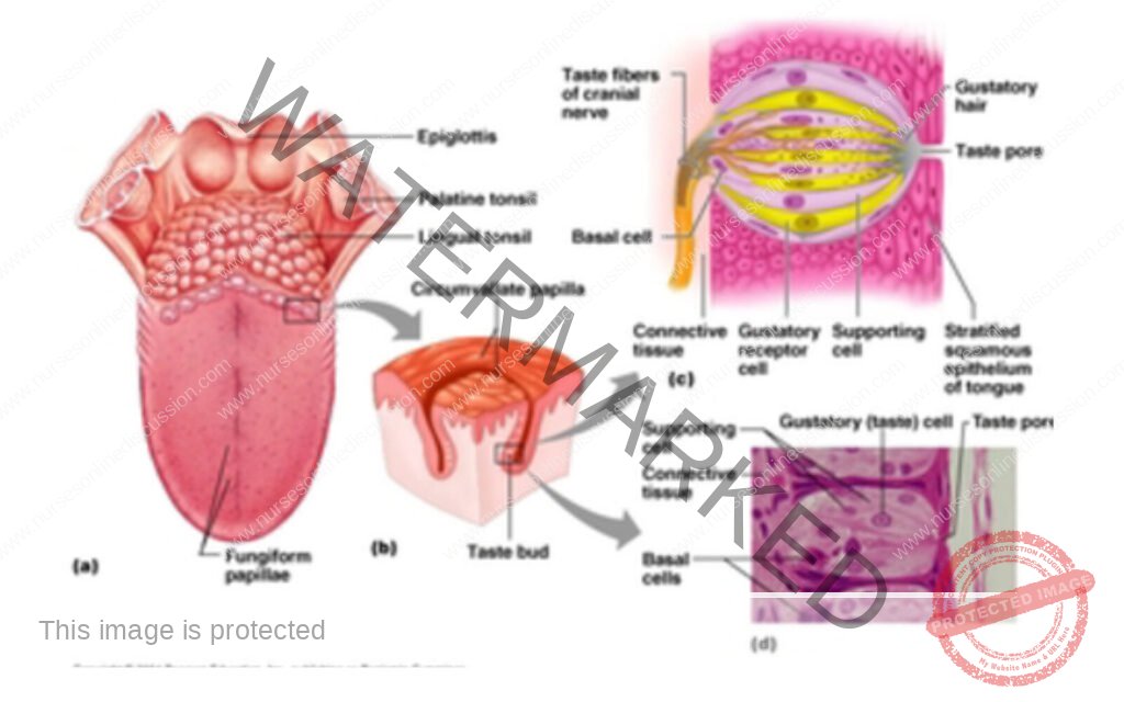
Taste buds are oval bodies found in projections of the tongue mucosa called the papillae which give the tongue surface a slightly abrasive feel. These papillae are of three major types i.e. filiform, fungiform and circumvallate. Only the latter two types house taste buds. The mush room shaped fungiform papillae are scattered all over the tongue surface but are most abundant on its tip and along the sides. The round circumvalet are the largest and least numerous of the papillae. 7-12 of these papillae form a V at the back of the tongue. Taste buds are located on the inner walls of the circumvalet papillae and on top of the fungiform papillae.
Each taste bud is a globular structure consisting of 40 to 60 epithelial cells of three major types i.e. supporting cells, taste cells and basal cells. Supporting cells form the bulk of the taste bud and surround receptor cells called taste or gustatory cells.
Micro villi called gustatory hair protrude from the tips of the taste cells through a taste pore to the surface of the bud, where they are bathed by saliva. Apparently, the gustatory hairs are the sensitive portions (receptor membranes) of the taste bud cells. Coiling intimately around the taste cells are sensory dendrites that represent the initial part of the gustatory pathway to the brain. Taste cells are shed every week to ten days and are replaced by new sensory cells derived from the division of basal cells.
HEARING SENSATION
The hearing organ the ear has external, middle and inner sections. The ear is also responsible for provision of the sense of equilibrium.
ANATOMY OF THE TASTE BUDS
External Ear
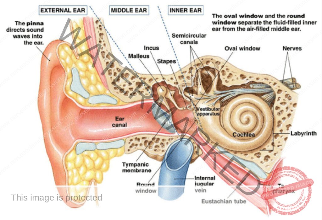
This includes all structures that face outside. These include the pinna (auricle), an S-shaped tube the external acoustic meatus (external auditory canal), typmpanic membrane (ear drum).
The external acoustic meatus passes into the temporal bone. It is lined with skin hairs and modified sweat glands called ceruminous gland that secrete wax. The hair and wax keep large foreign objects such as insects out of the ear.
The auricles of the ears help collect sound waves travelling in the air and direct them into the external acoustic meatus. After entering the meatus, sound waves pass to the end of the tube and alter the pressure on the tympanic membrane. The tympanic membrane is a semitransparent membrane covered by a thin layer of skin on its outer surface and by mucous membrane on the inside. The tympanic membrane moves back and forth in response the sound waves, reproducing the vibrations of the sound wave source.
Middle Ear
The middle ear or the tympanic cavity is an air-filled space in the temporal bone that separate the external and internal ears. It is bound by the tympanic membrane laterally and the inner ear medially and contains three bones called auditory ossicles.
The three auditory ossicles, the malleus, incus and stapes are attached to the wall of the tympanic cavity by tiny ligaments and are covered by mucous membrane. There functions include;
- They bridge the tympanic membrane and the inner ear, transmitting vibrations between the 2 parts.
- They for a lever system which aids in amplifying the vibrations as they pass from the tympanic membrane to the oval window.
Vibrations are then transmitted to the oval widow. This causes the movement of the fluid with in the inner ear. These vibrations of the fluid stimulate the hearing receptors. The round window is sealed by the secondary tympanic membrane and it vibrates with opposite phases to vibrations entering the inner ear through the oval window. The auditory tube connects the middle ear to the throat. It helps maintain equal air pressure on both sides of the tympanic membrane, which is necessary for normal hearing.
Inner Ear
This the most inner and complex part of the ear. It is mainly responsible for sound detection and balance. It consists of the bony labyrinth, cochlea that functions for hearing and three semicircular canals that provide a sense of equilibrium and vestibular system which houses membranous structures for hearing and equilibrium.
The bony labyrinth or osseous labyrinth is the network of passages with bony walls lined with periosteum. The membranous labyrinth runs inside of the bony labyrinth. There is a layer of perilymph fluid between them.
The cochlear is has 3 chambers which include the scala vestibuli, scala tympani, scala media. It is filled with a watery liquid, perilymph which moves in response to the vibrations coming from the middles ear via the oval window. As the fluid moves, the cochlear partition (basal membrane and organ of Corti) move thousands of hair cells and convert that motion into electrical signals that are then carried by the auditory nerve for interpretation in the brain.
SENSE OF SIGHT
A number of accessory organs assist the visual receptors which are in the eyes. These include the eyelids and lacrimal apparatus that help protect the eye and set of extrinsic muscles that move them.
Eyelids
It’s composed of for layers ie the skin, muscle, connective tissue and conjunctiva. The skin of the eyelid which is the thinnest skin of the body covers the lid`s outer surface and fuses with its inner lining near the margin of the lid. The muscles that move the eyelid include the orbicularis oculi and the levator palpebrae superioris. These act as sphincters that close the lids when it contracts.
Conjuctiva
This a mucous membrane that lines the inner surface of the eyelids and folds back to cover the anterior surface of the eyeball, except for its central portion the cornea.
Lacrimal Apparatus
This consist of the lacrimal gland which secret tears and a series of ducts which carry the tears into the nosal cavity. The gland is located in the orbit, superior and lateral to the eye.
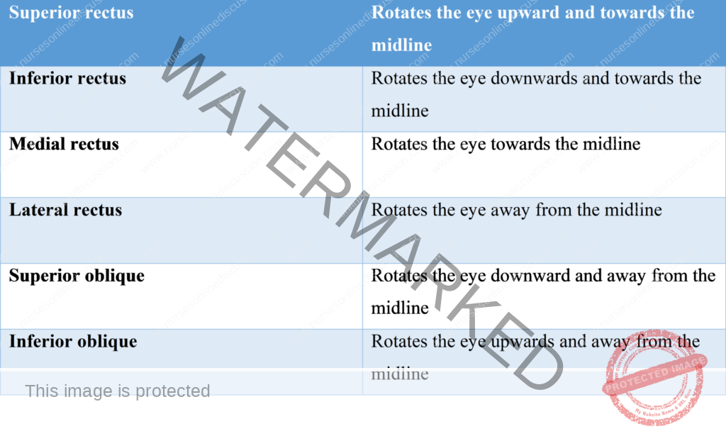
Extrinsic Muscles
These arise from the bones of the orbit and are inserted by broad tendons on the eye`s tough outer surface. Below are muscles that move the eye;
Structure of the Eye

The eye is a hollow spherical structure about 2.5cm. Its walls have 3 distinct layers which include; an outer fibrous tunic, middle vascular tunic and an inner nervous tunic.
The outer tunic
- It’s composed of the cornea, sclera
- The cornea is composed of largely connective tissue and a thin surface of epithelium. It acts as the window of the eye and help focus entering light rays.
- The sclera, the white portion of the eye makes up the posterior five-sixths of the outer tunic. The sclera protects the eye and is an attachment point for extrinsic muscles.
The Middle Tunic
- This layer includes the choroid coat, the ciliary body, lens, iris, pupil and aqueous humor
- The choroid coat joins loosely to the sclera. It contains blood vessels that nourish surrounding tissues. It also contains numerous melanocytes that give it a brownish black appearance. The melanin also helps absorb excess light and helps keep the inside of the eye dark.
- The ciliary body is the thickest part of the middle tunic. Within the ciliary processes are many radiating folds called ciliary processes and ciliary muscles. It also has many suspensory ligaments that hold the transparent lens in position. The inner surface of the ciliary body produces a watery fluid called aqueous humor which fills the space between the cornea and the lens providing nutrients and maintaining shape of the front of the eye.
The lens. The lacks blood vessels and is composed of specialized epithelial cells. It refracts light rays on to the retina.
The iris is a thin diaphragm mostly composed of connective tissue and smooth muscle fibers. The smooth muscle fibers of the iris form 2 groups, a circular set and radial set. These muscles control the size of the pupil through which light passes. The circular muscles act as sphincters, when it contracts, the pupil gets smaller. When the radial muscles the diameter of the pupil increases and the intensity of light entering increases.
The Inner tunic
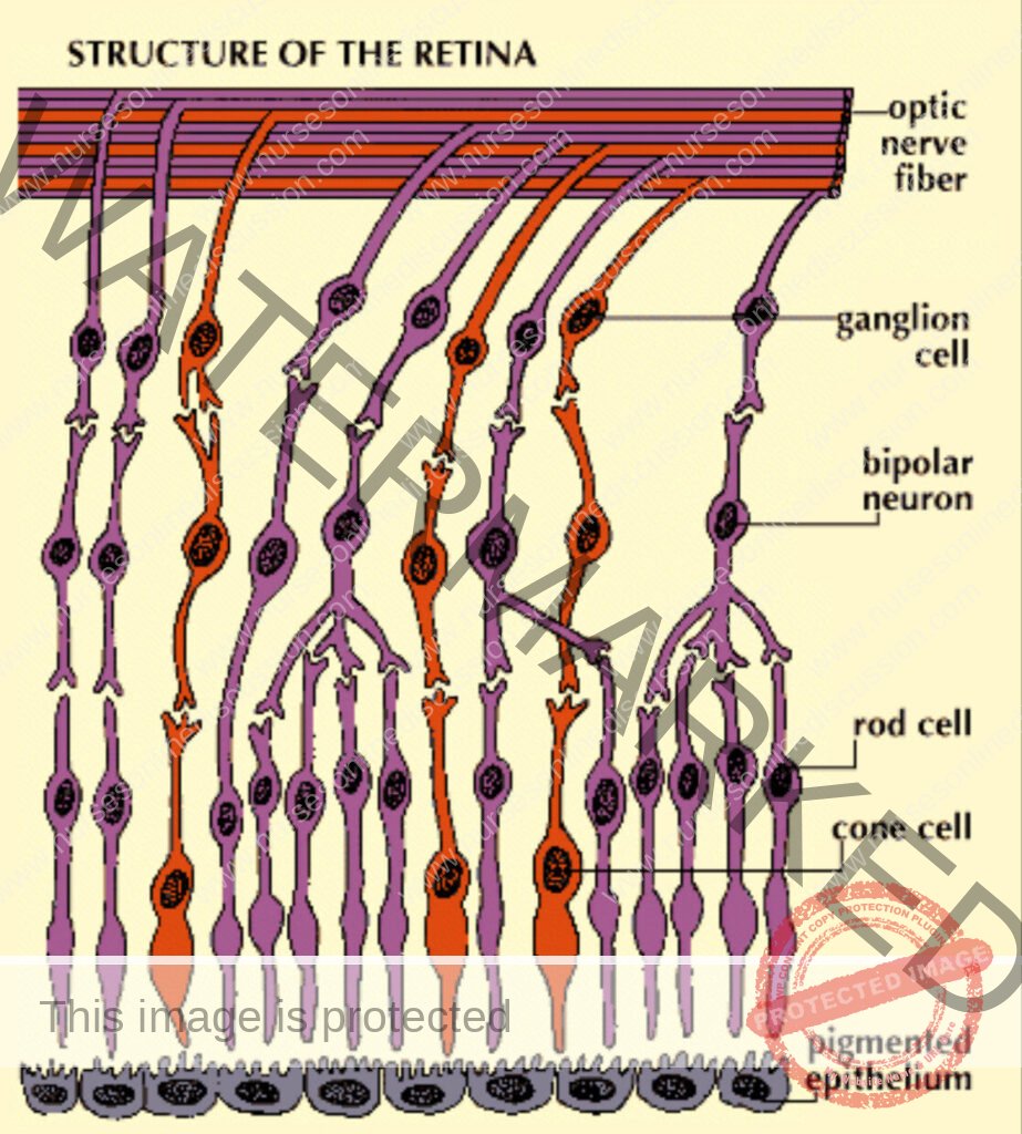
The inner tunics of the eye consist of the retina which contains visual receptor cells (photoreceptors). This nearly transparent sheet of tissue is continuous with the optic nerve in the back of the eye. It has distinct layers including pigmented epithelium, neurons, nerve fibers and limiting membranes.
There are five major groups of retinal neurons. The nerve fibers of the three groups; the receptor cells (rods and cones). Bipolar neurons and ganglion cells provide a direct pathway for impulses triggered in the receptors to the optic nerve then to the brain. The nerve fibers of the other two groups of retinal cells called the horizontal and amacrine cells, pass laterally between retinal cells. These modify the impulses transmitted on the fibers of the direct pathway. In the central region of the retina is a yellowish spot called the macula lutea, on its center is a depression called the fovea centralis, is the region of the retina that produces the sharpest vision. Medial to the fovea centralis is the optic disc where nerves leave the retina to the brain.
Summary
Light rays entering the eye must pass through the cornea, aqueous humor, lens, vitreous humor, and several layers of the retina before they reach the photoreceptors
We are a supportive platform dedicated to empowering student nurses and midwives through quality educational resources, career guidance, and a vibrant community. Join us to connect, learn, and grow in your healthcare journey
Quick Links
Our Courses
Legal / Policies
Get in Touch
(+256) 790 036 252
(+256) 748 324 644
Info@nursesonlinediscussion.com
Kampala ,Uganda
© 2026 Nurses online discussion. All Rights Reserved