Communicable Diseases
Subtopic:
Schistosomiasis

Schistosomiasis
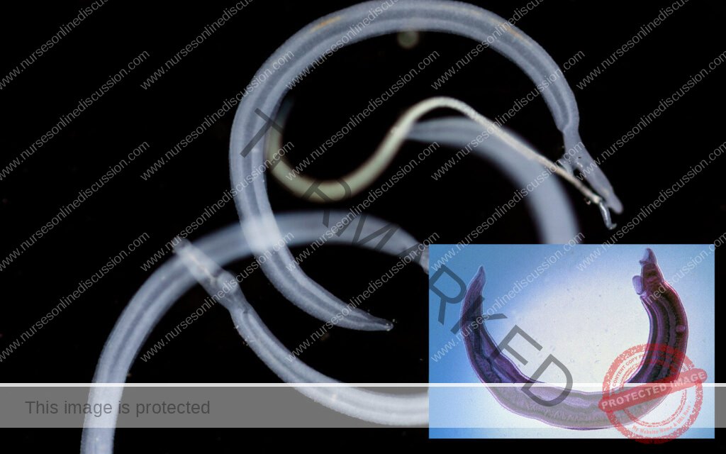
Schistosomiasis (also known as bilharzia or snail fever) is a disease of the large intestine and urinary tract caused by parasitic worms of the Schistosoma blood fluke.
It may infect the urinary tract or intestines.
Cause
- Schistosoma haematobium causes urinary schistisomiasis.
- Schistosomamansoni and Schistosoma intercalatum cause intestinal schistosomiasis.
- Schistosoma japonicum and Schistosoma mekongi cause Asian intestinal schistosomiasis.
- Avian schistosomiasis species cause swimmer’sitch.
Transmission
The disease is spread by contact with water that contains the parasites. These parasites are released from freshwater snails that have been infected.
The larvae form (cercariae) of schistsoma penetrates the skin from contaminated water.
Epidemiology
- Schistosomiasis affects almost 210 million people worldwide.
- An estimated 12,000 to 200,000 people die from it a year.
- The disease is most commonly found in Africa, Asia and South America.
- Around 700 million people, in more than 70 countries, live in areas where the disease is common.
- Schistosomiasis is second only to malaria, as a parasitic disease with the greatest
Signs and symptoms
- Above all, schistosomiasis is a chronic disease.
Schistosoma haematobium (urinary tract)
- Painless blood stained urine at the end of urination (haematuria)
- Frequency of urination (cystitis and fibrosis)
- Hydronephrosis, pyonephrosis, hypertension, uraemia.
- Fatigue
- Genital sores
Schistsoma mansoni (GIT)
- Abdominal pain
- Frequent stool with blood stained mucous
- Palpable liver (hepatomegally)
- Haematemesis
- Cough
- Diarrhea
- Fever
- Fatigue
Other symptoms
- Skin symptoms: At the start of infection, mild itching and a papular dermatitis of the feet and other parts after swimming in polluted streams containing cercariae.
Life cycle
Snails
The life cycles of all human schistosomes are similar.
- Parasite eggs are released into the environment from infected individuals, hatching on contact with fresh water to release the free-swimming miracidium.
- Miracidia infect freshwater snails by penetrating the snail’s foot.
- After infection, close to the site of penetration, the miracidium transforms into a primary (mother) sporocyst.
- Germ cells within the primary sporocyst will then begin dividing to produce secondary (daughter) sporocysts, which migrate to the snail’s hepatopancreas.
- Once at the hepatopancreas, germ cells within the secondary sporocyst begin to divide again, this time producing thousands of new parasites, known as cercariae, which are the larvae capable of infecting mammals.
Humans
- Penetration of the human skin occurs after the cercaria have attached to and explored the skin. The parasite secretes enzymes that break down the skin’s protein to enable penetration of the cercarial head through the skin.
- As the cercaria penetrates the skin it transforms into a migrating schistosomulum The newly transformed schistosomulum may remain in the skin for two days
- From here the schistosomulum travels to the lungs where it undergoes further developmental changes necessary for subsequent migration to the liver.
- 8 to 10days after penetration of the skin, the parasite migrates to the liver sinusoids. Young mansoni and S. japonicum worms develop an oral sucker after arriving at the liver, and it is during this period that the parasite begins to feed on red blood cells.
- The nearly-mature worms pair, and relocate to the mesenteric or rectal veins. haematobium schistosomula ultimately migrate from the liver to the bladder, ureters, and kidneys through the hemorrhoidal plexus.
- Parasites reach maturity in six to eight weeks, at which time they begin to produce eggs.
- Many of the eggs pass through the walls of the blood vessels, and through the intestinal wall, to be passed out of the body in feces.
- Schistosoma haematobium eggs pass through the ureteral or bladder wall and into the urine.
- Only mature eggs are capable of crossing into the digestive tract, possibly through the release of proteolytic enzymes, but also as a function of host immune response, which fosters local tissue ulceration.
- Worm pairs can live in the body for an average of four and a half years, but may persist up to twenty years.
Diagnosis
- History of staying in an endemic area.
- Urine examination (for schistosoma haematobium ova)
- Stool examination (for schistosoma mansoni ova)
- Bladder x-ray for calcification
- Ultrasound for urinary tract
- Detection of parasitic antigens by ELISA (blood sample).
- Tissue biopsy: (rectal biopsy (snip) for all species and biopsy of the bladder for haematobium) may demonstrate eggs when stool or urine examinations are negative.
Treatment
- Schistosomiasis is readily treated using a single oral dose of the drug Praziquantel annually, (40mg/kg).
Prevention
- Avoid defecating or urinating in or near water
- Avoid washing or stepping in contaminated water
- Effective treatment of cases
- When a village reports more than 50 percent of children have blood in their urine, everyone in the village receives treatment.
- Clear bushes around landing sites
- Eliminating the water-dwelling snails that are the natural reservoir of the disease.
- Copper sulfate and Niclosamide can be used for this purpose.
Get in Touch
(+256) 790 036 252
(+256) 748 324 644
Info@nursesonlinediscussion.com
Kampala ,Uganda
© 2025 Nurses online discussion. All Rights Reserved Design & Developed by Opensigma.co

