Conditions Affecting the Nervous System
Subtopic:
Applied Anatomy and Physiology of the Nervous System
Table of Contents

Learning Objectives
Describe the structural and functional divisions of the nervous system, including CNS and PNS components.
Identify major neuron structures and explain their roles in nerve impulse transmission.
Classify neuron types—sensory, motor, and interneurons—and outline their functions.
Recognize supporting neuroglial cells in CNS and PNS and summarize their physiological significance.
Explain the anatomy and physiology of the brain and spinal cord, including major regions and functions.
Discuss peripheral nerve organization, spinal nerve classification, and clinical concepts like dermatomes and referred pain.
Nervous System
The nervous system is a complex network of specialized cells and tissues that coordinates actions by transmitting signals to and from different parts of the body. It is responsible for detecting, interpreting, and responding to internal and external stimuli.
Anatomy of the Nervous System
The nervous system can be divided both structurally and functionally:
Structural Divisions:
Central Nervous System (CNS): Consists of the brain and spinal cord, which develop from the neural tube during embryonic development. The CNS acts as the control center, processing information and generating responses.
Peripheral Nervous System (PNS): Composed of all nervous structures outside the CNS. These structures connect the CNS to the rest of the body. The PNS develops from neural crest cells and CNS outgrowths. It includes:
Cranial Nerves: Nerves that emerge directly from the brain.
Spinal Nerves: Nerves that emerge from the spinal cord.
Visceral Nerves and Plexuses: Nerves that innervate internal organs.
Enteric Nervous System: A specialized network of nerves within the walls of the digestive tract.
Functional Divisions:
Somatic Nervous System: Controls voluntary movements and receives sensory information from the external environment. It primarily innervates structures derived from somites during development, such as skin and skeletal muscle.
Visceral Nervous System (Autonomic Nervous System): Controls involuntary functions of internal organs, smooth muscle, and glands. It primarily monitors and regulates the internal environment.
The Neuron: Structure and Function
The neuron is the fundamental functional unit of the nervous system. It is a specialized cell designed to transmit information via electrochemical signals. Each neuron consists of a cell body and an axon that terminates in a synapse, a specialized junction for communication with other neurons or target cells (e.g., muscles, glands).
Structure of a Neuron:

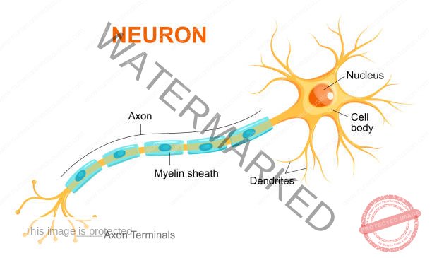
Functions of Neuron Structures
A typical neuron is composed of several key structures, each with a specific role in nerve impulse transmission:
Cell Body (Soma): The central part of the neuron, containing the nucleus and cytoplasm.
Functions:
Houses the nucleus, which contains the genetic material (DNA) and controls the neuron’s activities.
Acts as the neuron’s metabolic center, producing proteins and neurotransmitters necessary for neuronal function.
Integrates incoming signals from the dendrites.
Dendrites: Branch-like extensions that receive signals (stimuli) from other neurons or sensory receptors and transmit them towards the cell body.
Axon: A long, slender fiber that conducts nerve impulses away from the cell body towards other neurons, muscles, or glands. It forms the conductive region of the neuron.
Myelin Sheath: A fatty, insulating layer that surrounds many axons, particularly in the peripheral nervous system.
Function:
Increases the speed of nerve impulse transmission.
Provides insulation, preventing signal loss.
Schwann Cells: Specialized glial cells that produce the myelin sheath in the peripheral nervous system (PNS). They also play a role in axon maintenance and regeneration.
Nodes of Ranvier: Gaps in the myelin sheath along the axon.
Function: Allow for faster nerve impulse conduction through a process called saltatory conduction, where the impulse “jumps” from one node to the next.
Axon Terminals: The branched endings of an axon.
Function: Form synapses (junctions) with other neurons or target cells, where neurotransmitters are released to transmit signals.
Types of Neurons
Neurons can be broadly classified into three main types based on their function:
Sensory Neurons (Afferent Neurons): Transmit sensory information from sensory receptors (e.g., in the skin, eyes, ears) towards the central nervous system (CNS).
Motor Neurons (Efferent Neurons): Transmit signals from the CNS away to muscles and glands, causing movement or secretion.
Interneurons (Association Neurons): Located within the CNS, they connect sensory and motor neurons, processing and integrating information. They form complex neural circuits.

Other Cells of the Nervous System
Neuroglia (Glial Cells): These are non-neuronal cells that provide support, protection, and nourishment to neurons. They play crucial roles in maintaining the proper functioning of the nervous system. Examples include astrocytes, oligodendrocytes, microglia, and ependymal cells in the CNS, and Schwann cells and satellite cells in the PNS.
- Satellite Cells (PNS):
Location: Surround neuron cell bodies within ganglia (clusters of neuron cell bodies) in the peripheral nervous system (PNS).
Function: Provide structural support, regulate the chemical environment around neurons, and insulate the ganglia.
Ependymal Cells (CNS):
Location: Line the ventricles of the brain and the central canal of the spinal cord.
Functions:
Produce cerebrospinal fluid (CSF).
Form the choroid plexuses, specialized structures within the ventricles that are the primary sites of CSF production.
Help circulate CSF with cilia.
Oligodendrocytes (CNS):
Location: Central nervous system (CNS).
Function: Form the myelin sheath around axons in the CNS, providing insulation and increasing the speed of nerve impulse transmission. They are functionally analogous to Schwann cells in the PNS.
Astrocytes (CNS):
Location: Central nervous system (CNS).
Functions:
Provide structural support to neurons.
Anchor neurons to blood vessels.
Regulate the chemical environment around neurons.
Contribute to the formation of the blood-brain barrier, a protective barrier that regulates the passage of substances from the blood to the brain.
Star-shaped in appearance.
Microglia (CNS):
Location: Central nervous system (CNS).
Function: Act as the immune cells of the CNS, performing phagocytosis (engulfing and removing cellular debris, pathogens, and damaged tissue). They are derived from monocytes.
CENTRAL NERVOUS SYSTEM (CNS
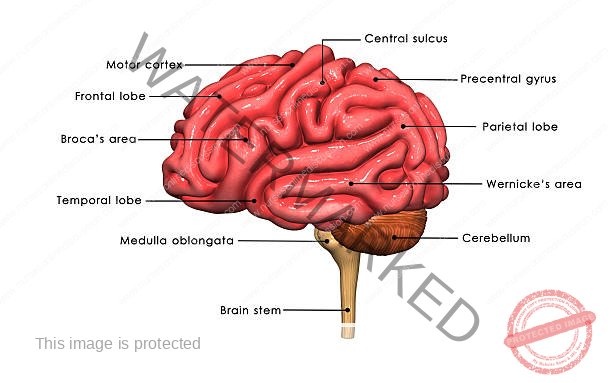
The Brain: Anatomy
The brain, the most complex organ in the body, is the central processing unit of the nervous system. It is divided into several major parts:
Cerebrum:
The largest part of the brain, responsible for higher-level functions like thought, memory, and voluntary movement.
Divided into two cerebral hemispheres (left and right) by the longitudinal cerebral fissure.
The corpus callosum, a large bundle of white matter fibers, connects the two hemispheres deep within the fissure, allowing for communication between them.
Structure:
Cerebral Cortex: The outer layer, composed of gray matter (neuron cell bodies), responsible for higher-level cognitive functions.
White Matter: The inner layer, composed of myelinated axons, responsible for communication between different brain regions.
Lobes: Each hemisphere is further divided into four lobes:
Frontal Lobe: Involved in planning, decision-making, problem-solving, voluntary movement, and speech.
Parietal Lobe: Processes sensory information, including touch, temperature, pain, and pressure; also involved in spatial awareness.
Temporal Lobe: Processes auditory information, memory, and language comprehension.
Occipital Lobe: Processes visual information.
Cerebellum:
Located at the back of the brain, below the occipital lobe of the cerebrum and behind the brainstem, occupying the posterior cranial fossa.
Has an oval shape with two hemispheres connected by a central portion called the vermis.
Structure: Similar to the cerebrum, it has an outer layer of gray matter and an inner core of white matter.
Brainstem:
Connects the cerebrum and cerebellum to the spinal cord.
Composed of the midbrain, pons, and medulla oblongata.
Midbrain:
Located below the cerebrum and above the pons, surrounding the cerebral aqueduct.
Contains nuclei (clusters of neuron cell bodies) and nerve fibers that connect the cerebrum to lower parts of the brain and the spinal cord.
Pons:
Located in front of the cerebellum, below the midbrain, and above the medulla oblongata.
Contains nuclei and nerve fibers, including some cranial nerve nuclei.
Medulla Oblongata:
Extends from the pons and is continuous with the spinal cord.
Contains vital regulatory centers for breathing, heart rate, and blood pressure.
Composed of central gray matter surrounded by outer white matter tracts connecting to the spinal cord.
Diencephalon:
Located deep within the brain, connecting the cerebrum and the midbrain.
Includes structures surrounding the third ventricle, the pineal gland, and the optic chiasm.
Thalamus:
Two masses of gray and white matter located below the corpus callosum, on either side of the third ventricle.
Acts as a major relay station for sensory information.
Hypothalamus:
Located below and in front of the thalamus.
Connected to the pituitary gland.
Contains several nuclei that regulate various bodily functions.
Physiology of the Brain
Cerebral Cortex:
Higher Mental Processes: Responsible for complex cognitive functions, including learning, memory, reasoning, and language.
Sensory Perception: Processes sensory input from the body, including pain, temperature, touch, sight, hearing, taste, and smell.
Motor Control: Initiates and controls voluntary muscle contractions.
Cerebellum:
Coordination: Coordinates voluntary muscle movements, ensuring smooth and precise actions.
Posture and Balance: Maintains posture and balance.
Motor Learning: Involved in learning and refining motor skills.
Language Processing: Plays a role in some aspects of language processing.
Thalamus:
Sensory Relay: Relays sensory impulses from most parts of the body to the cerebral cortex.
Memory: Involved in some aspects of memory.
Hypothalamus:
Autonomic Control: Regulates the autonomic nervous system (involuntary functions like heart rate, digestion, and breathing).
Homeostasis: Controls essential bodily functions, including appetite, thirst, body temperature, and water balance.
Emotional Responses: Influences emotional reactions and behavior.
Sexual Behavior: Regulates sexual behavior and responses.
Sleep-Wake Cycles: Regulates the sleep-wake cycle through the influence of melatonin from the nearby pineal gland.
Endocrine Control: Secretes hormones like antidiuretic hormone (ADH) and oxytocin and controls the release of hormones from the pituitary gland.
Brainstem:
Midbrain: Contains nuclei that serve as relay stations for ascending and descending nerve fibers, connecting the cerebrum with lower brain regions and the spinal cord.
Pons: Works with the medulla to regulate breathing and serves as a relay station for some cranial nerves.
Medulla Oblongata:
Vital Centers: Contains the respiratory center (controls breathing), cardiovascular center (regulates heart rate and blood pressure), and reflex centers (controls vomiting, coughing, sneezing, and swallowing).
Decussation of Pyramids: The site where motor fibers from the cerebral cortex (corticospinal tracts) cross over to the opposite side of the body. This is why the left side of the brain controls the right side of the body, and vice versa.
Anatomy of the Spinal Cord
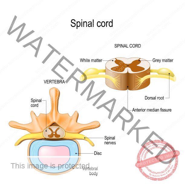
The spinal cord, a crucial component of the central nervous system, is a long, cylindrical structure that extends from the brainstem down through the vertebral canal. It serves as the main pathway for communication between the brain and the rest of the body.
Structure:
Shape: Roughly cylindrical, appearing circular to oval in cross-section.
Central Canal: A small, cerebrospinal fluid (CSF)-filled canal runs through the center of the spinal cord.
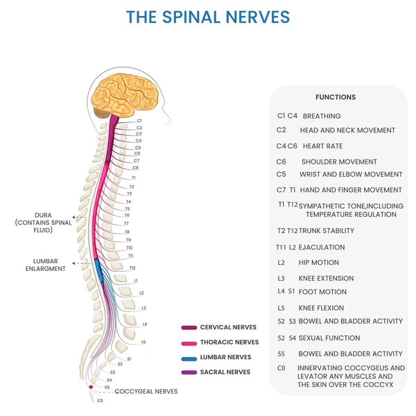
Spinal Nerves: 31 pairs of spinal nerves emerge from the spinal cord, each with two roots:
Dorsal Root (Sensory): Carries sensory information from the body to the spinal cord (afferent fibers).
Ventral Root (Motor): Carries motor commands from the spinal cord to muscles and glands (efferent fibers).
Extent:
Adults: Extends from the foramen magnum (the opening at the base of the skull) to approximately the level of the intervertebral disc between the first and second lumbar vertebrae (L1-L2). However, it can terminate as high as the twelfth thoracic vertebra (T12) or as low as the disc between the second and third lumbar vertebrae (L2-L3).
Neonates: Extends lower, typically to the third lumbar vertebra (L3) or even the fourth (L4).
Conus Medullaris: The tapered, cone-shaped distal end of the spinal cord.
Filum Terminale: A thin filament of connective tissue that extends inferiorly from the tip of the conus medullaris, anchoring the spinal cord to the coccyx.
Enlargements: Two major swellings along the spinal cord:
Cervical Enlargement: Located in the cervical region (C5-T1), corresponding to the origin of spinal nerves that innervate the upper limbs.
Lumbosacral Enlargement: Located in the lumbar region (L1-S3), corresponding to the origin of spinal nerves that innervate the lower limbs.
Internal Structure:
Gray Matter: The central, H-shaped region, rich in neuron cell bodies, dendrites, and unmyelinated axons. It is involved in processing and integrating information.
White Matter: Surrounds the gray matter and is composed primarily of myelinated axons, forming tracts that carry information up and down the spinal cord (ascending and descending tracts) and to and from the brain.
Physiology of the Spinal Cord
The spinal cord serves as a vital communication link between the brain and the peripheral nervous system.
Conduction Pathways: Tracts within the white matter of the spinal cord transmit sensory information up to the brain (ascending tracts) and motor commands down from the brain to muscles and glands (descending tracts).
Reflex Integration: The gray matter of the spinal cord integrates reflexes, which are rapid, involuntary responses to stimuli. These reflexes occur at the level of the spinal cord without immediate input from the brain.
The Meninges
The meninges are three layers of connective tissue membranes that surround, protect, and support the brain and spinal cord within the cranial cavity and vertebral canal.
Dura Mater:
The outermost, toughest, and most fibrous layer.
Continuous with the cranial dura mater at the foramen magnum.
In the cranial cavity, a layer of the dura mater is fused to the skull bones, serving as the periosteum.
In the vertebral canal, the spinal dura mater is separated from the vertebrae by an epidural space.
The dural sac narrows at the lower border of vertebra S2 and forms a sheath for the filum terminale, which anchors to the coccyx.
Arachnoid Mater:
A thin, delicate, web-like membrane located beneath the dura mater.
Not directly attached to the dura but held against it by the pressure of the CSF.
Separated from the pia mater by the subarachnoid space, which contains cerebrospinal fluid (CSF).
Ends at the level of vertebra S2.
Pia Mater:
The innermost, thin, and vascular membrane that adheres closely to the surface of the brain and spinal cord.
Extends into the anterior median fissure of the spinal cord.
Forms sleeve-like coverings around the spinal nerve roots as they cross the subarachnoid space.
Denticulate Ligaments: Paired, longitudinal extensions of pia mater that anchor the spinal cord laterally to the arachnoid and dura mater.
Clinical Significance of Lumbar Region:
The subarachnoid space can be safely accessed in the lower lumbar region (below L2 in adults) without risking spinal cord injury.
The L4 vertebral spinous process can be located by drawing a horizontal line between the highest points of the iliac crests.
In the lumbar region, the palpable tips of the spinous processes are at the same level as their corresponding vertebral bodies.
The subarachnoid space can be accessed between L3-L4 or L4-L5 for a lumbar puncture.
PERIPHERAL NERVOUS SYSTEM (PNS)
The peripheral nervous system (PNS) consists of all the nervous tissue outside the central nervous system (CNS). It connects the CNS to the limbs and organs, essentially serving as a communication relay going back and forth between the brain and spinal cord with the rest of the body.
Cranial Nerves
there are 12 pairs of cranial nerves.
Olfactory Nerve (CN I):
Originates from olfactory receptors in the nasal mucosa.
Function: Sense of smell.
Clinical Note: Loss of smell (anosmia) can be caused by head injuries or tumors affecting the olfactory groove.
Assessment: Tested by assessing the ability to smell different odors through each nostril.
The remaining 11 nerves will be explained in the next refinement.
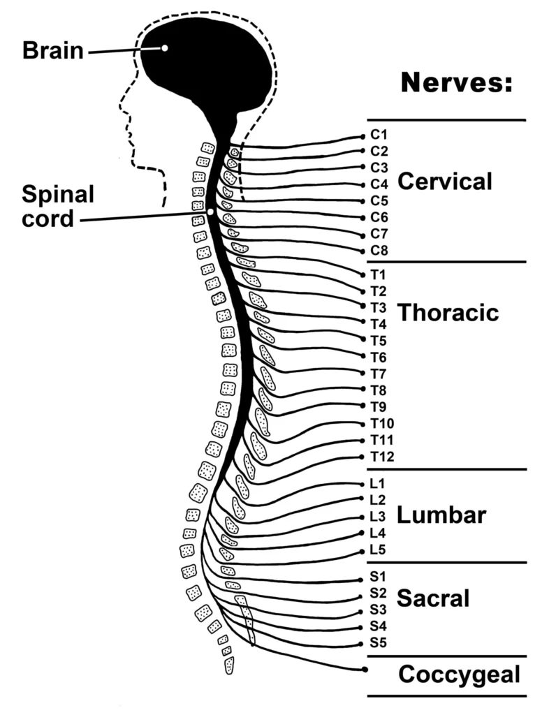
Spinal Nerves
Spinal nerves are mixed nerves, containing both sensory (afferent) and motor (efferent) fibers.
There are 31 pairs of spinal nerves, named and numbered according to the region of the vertebral column from which they emerge:
8 cervical nerves (C1-C8)
12 thoracic nerves (T1-T12)
5 lumbar nerves (L1-L5)
5 sacral nerves (S1-S5)
1 coccygeal nerve (Co1)
Nerve C1 emerges between the skull and the atlas (first cervical vertebra). All other spinal nerves emerge below their corresponding vertebrae (or former vertebrae in the case of the sacral nerves).
Plexus: A network formed by the interconnection of nerve fibers, where new combinations of fibers emerge as named peripheral nerves.
Dermatomes: Specific areas of skin innervated by sensory fibers from a single spinal nerve. Dermatome maps are useful in diagnosing the origin of pain, numbness, or tingling when these symptoms are caused by spinal cord or nerve root compression
Referred Pain
Referred pain occurs when pain from an internal organ is felt in a different area of the body surface. This happens because sensory fibers from the organ and the skin share the same spinal nerve root. The brain may misinterpret the organ pain as coming from the skin. For example, heart pain can be felt in the left arm, and diaphragm pain can be felt in the left shoulder.
Myotomes
A myotome is a group of muscles primarily innervated by a single spinal nerve. Since most muscles receive innervation from multiple spinal levels, myotomes are assessed by testing joint movements or muscle groups. This helps identify the spinal nerve or spinal cord level that might be affected.
Join Our WhatsApp Groups!
Are you a nursing or midwifery student looking for a space to connect, ask questions, share notes, and learn from peers?
Join our WhatsApp discussion groups today!
Join NowRelated Topics
Conditions Affecting the Nervous System
- Applied Anatomy and Physiology of the Nervous System
- Trigeminal Neuralgia
- Bell’s Palsy
- Parkinson’s Disease
- Spinal Cord Compression
- Transverse Myelitis
Medical Conditions Affecting the Endocrine System
- Applied Anatomy and Physiology of the Endocrine System
- Acromegaly/Gigantism (Hyperpituitarism)
- Dwarfism (Panhypopituitarism)
- Addison’s Disease (Adrenal Insufficiency)
- Pheochromocytoma
- Cushing’s Syndrome
- Hyperaldosteronism
- Thyrotoxicosis
- Diabetes Mellitus
Medical Diseases Affecting the Renal System
- Anatomy and Physiology of the Renal System
- Renal Disorders
- Urinary Tract Infections (UTIs)
- Cystitis
- Renal Failure (Acute and Chronic)Nephrotic Syndrome
- Polycystic Kidney Disease (PKD)
- Kidney Stones (Nephrolithiasis)
Conditions of the Lymphatic System
- Anatomy and Physiology of the Lymphatic System
- Lymphedema
- Lymphangitis and Lymphadenitis
- Hodgkin’s Disease
Conditions of the Musculo-Skeletal System
- Anatomy and Physiology of the Musculo-Skeletal System
- Tendonitis
- Rheumatoid Arthritis
- Osteoarthritis
- Gout
- Bursitis
- Ankylosing Spondylitis
- Osteoporosis
- Paget’s Disease
Skin Conditions (Dermatology)
We are a supportive platform dedicated to empowering student nurses and midwives through quality educational resources, career guidance, and a vibrant community. Join us to connect, learn, and grow in your healthcare journey
Quick Links
Our Courses
Legal / Policies
Get in Touch
(+256) 790 036 252
(+256) 748 324 644
Info@nursesonlinediscussion.com
Kampala ,Uganda
© 2026 Nurses online discussion. All Rights Reserved