Obstetric Anatomy and Physiology
Fertilization and Implantation
Table of Contents
Definition
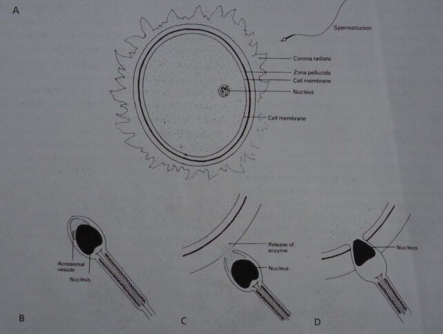
Fertilisation is the union of an Ovum and a spermatozoon. It usually occurs at the Ampulla of the fallopian tube approximately $36-48$ hours after ovulation.
The Zygote
The cell formed from this union is called a Zygote and it contains 46 chromosomes in the nucleus.
Individual sex is determined by the $\text{X}$ and $\text{Y}$ chromosomes: $\text{X}$ = Female; $\text{Y}$ = Male.
DEVELOPMENT OF THE ZYGOTE AND BLASTOCYST
Early Zygote Development
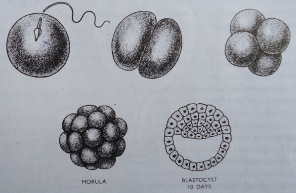
The zygote travels towards the uterus, aided by the ciliary and peristaltic movement of the fallopian tube. It is nourished by secretions in the tube and food previously stored in the ovum. During this 3-day journey, the zygote divides repeatedly to form a solid mass of cells called the Morula.
Diagrammatic representation of the fusion of the ovum and the spermatozoon
DEVELOPMENT OF THE FERTILIZED OVUM
The Blastocyst
Later, a cavity develops at one end of the morula as fluid collects, and this structure is known as the Blastocyst. By the 4th to 5th day, the blastocyst arrives at the uterus.
The blastocyst structure consists of:
Trophoblast: The outer layer of cells.
Inner Cell Mass: Layers of cells collected at one end of the pole, which will form the embryo.
Body Stock: The cells connecting the inner cell mass to the trophoblast.
Formation of the Decidua
As a result of conception, the Corpus Luteum continues to grow and produces more Progesterone under the influence of Human Gonadotrophin Hormones.
This promotes the growth and development of the secretory Endometrium into the Decidua.
The glands in the Endometrium dilate, become more torturous, and fill with secretions.
The Endometrium becomes soft, spongy, and highly vascularized to allow for embedment.
Embedment (Implantation)
The blastocyst usually rests on the decidua of the fundus of the uterus.
The trophoblast secretes an enzyme which erodes the decidua and some blood vessels.
This allows the blastocyst to embed in the decidua with the inner cell mass in the lead.
After embedment, the decidua is named based on its location:
Decidua Basalis: The decidua directly under the blastocyst (where the placenta will form).
Decidua Capsularis: The decidua surrounding and covering the blastocyst.
Decidua Vera: The undisturbed decidua lining the rest of the uterus.
THE TROPHOBLAST
Trophoblast Structure and Function
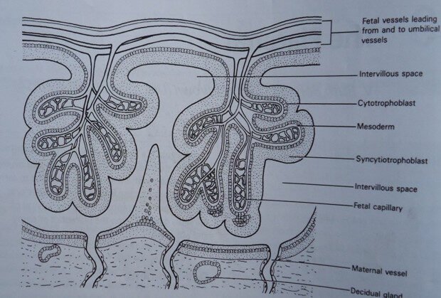
The trophoblast cells multiply, forming finger-like projections called villi on the surface of the blastocyst. These villi invade the decidua and maternal blood vessels so that each projection is surrounded by maternal blood.
The trophoblastic cell layers are:
Syncytium or Syncytiotrophoblast: The outer layer.
Cytotrophoblast: The inner layer.
Mesoderm: The layer below (loose connective tissue).
Syncytiotrophoblast
This outer layer is composed of a nucleated layer of protoplasm. It is capable of breaking down tissue (eroding the decidua) and blood vessel walls, making nutrients and oxygen in the maternal blood accessible to the developing embryo.
Cytotrophoblast
This inner layer secretes the Human Gonadotrophic Hormone (H.C.G.), which stimulates the corpus luteum to continue producing Oestrogen and Progesterone. This maintains the secretory phase and prevents shedding (menstruation) from taking place, thus maintaining the pregnancy.
The Mesoderm
The mesoderm consists of loose connective tissue, similar to that of the inner cell mass, and the two are continuous at the point where they join the body stalk.
THE INNER CELL MASS
Three Germ Layers
The Inner Cell Mass forms the fetus itself and develops into the three primary germ layers:
Ectoderm: Mainly forms the skin, hair, nails, the lens of the eyes, enamel of the teeth, and the nervous system.
Mesoderm: Forms bones and muscles, the heart and blood vessels (including those in the placenta), kidneys, ovaries, testes, and other internal organ muscles.
Endoderm: Forms the alimentary tract, liver, pancreas, lungs, and thyroid glands.
Summary of Blastocyst Derivatives: The Trophoblasts form the chorion and placenta, while the Inner Cell Mass forms the fetus, amnion, and umbilical cord.
The Amniotic Cavity
This cavity lies over the side of the ectoderm. It is filled with amniotic fluid and gradually enlarges, folding around the embryo to enclose it. The amnion membrane forms from its lining and swells out into the chorionic cavity (which eventually dissolves as the amnion and chorion fuse).
The Yolk Sac
This lies on the side of the endoderm. It provides early nourishment before the placenta takes over. Part of the yolk sac contributes to the formation of the primitive gut. The remainder resembles a balloon, floating in front of the embryo until it atrophies and becomes trapped under the amnion on the surface of the placenta.
DEVELOPMENT OF THE PLACENTA
Chorionic Development
Three weeks after fertilization, one portion of the primitive chorion, next to the Decidua Basalis, outgrows the other portion, which later atrophies.
Chorion Frondosum: The portion that outgrows the other (will form the placenta).
Chorion Laeve: The portion that atrophies (will form the smooth chorion membrane).
Villi and Placental Structure
Each villus on the chorion frondosum branches into tree-like projections. Each branch has three layers, and the intervillous spaces are filled with maternal blood.
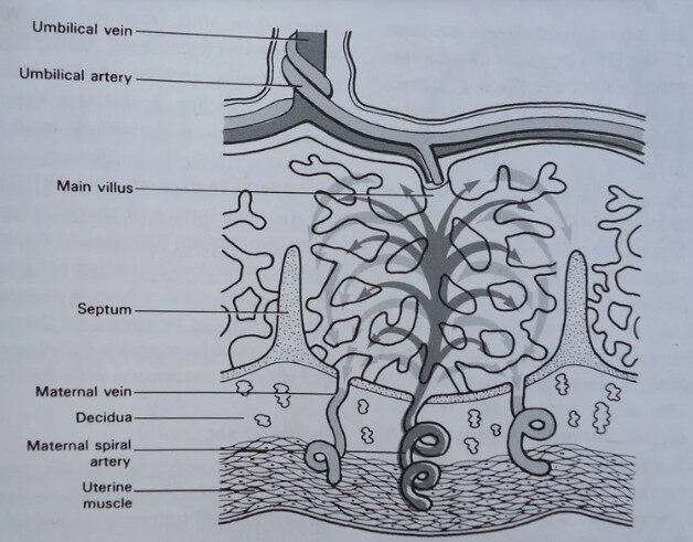
Anchoring Villi: Some larger branches penetrate deep into the decidua to anchor the placenta.
Nutritive Villi: The smaller villi which float into the maternal blood and facilitate exchange.
The collection of villi of the chorion frondosum forms the Placenta. The placenta is fully developed at the $12^{th}$ week of pregnancy.
Diagram showing chorionic villi
Placental Circulation
The villi allow for the exchange of nutrients, oxygen, and waste between the maternal blood (surrounding the villi) and the fetal blood (inside the villi) without the two bloodstreams ever mixing.
Circulation though the placenta
Join Our WhatsApp Groups!
Are you a nursing or midwifery student looking for a space to connect, ask questions, share notes, and learn from peers?
Join our WhatsApp discussion groups today!
Join NowGet in Touch
(+256) 790 036 252
(+256) 748 324 644
Info@nursesonlinediscussion.com
Kampala ,Uganda
© 2025 Nurses online discussion. All Rights Reserved Design & Developed by Opensigma.co