Bone Conditions in Children
Subtopic:
Fractures
Fractures represent a disruption, either complete or incomplete, in the continuous structure of a bone.
A fracture occurs when the structural integrity of bone tissue is compromised due to the application of excessive or unusual force.
Essentially, a fracture is a break in a bone that happens when the force applied exceeds the bone’s capacity to withstand it.
Fractures are also commonly referred to as broken bones.
Common Childhood Fractures
Bones in the arm are more frequently fractured in children compared to other bones.
Collarbone or shoulder fractures: Breaks affecting the clavicle or scapula.
Elbow fractures: Fractures occurring at the joint of the upper and lower arm.
Forearm, wrist, or hand fracture: Breaks in the radius, ulna, carpal, or metacarpal bones.
Leg, foot, or ankle fracture: Fractures of the tibia, fibula, tarsal, metatarsal, or phalangeal bones.
Causes of Fractures
Direct Force: The fracture happens directly at the point where the impact occurs.
Torsion: The fracture occurs away from the point of impact, often due to a twisting force. For instance, a twisted foot can cause a break in the leg bones.
Violent Contractions: Powerful muscle contractions can generate enough force to fracture a bone. An example is fracturing the humerus while forcibly throwing something, or during strong muscle spasms like in tetanus.
Disease Processes: Certain conditions can weaken bone structure, making them more susceptible to fractures. Examples include osteoporosis, malnutrition that weakens bones, and bone tumors that compromise bone integrity.
Risks for fractures
Sporting accidents: Injuries sustained during athletic activities.
Falls from heights: Accidents involving a drop from an elevated position.
Bike and car accidents: Trauma resulting from collisions involving vehicles.
Poor nutrition; a diet low in calcium: Insufficient calcium intake can lead to weakened bones.
Associated events following fractures
When a bone breaks, surrounding tissues are inevitably affected, leading to:
Soft tissue edema: Swelling in the tissues around the fracture site.
Joint dislocation: Displacement of bones within a joint.
Ruptured tendons: Tearing of the fibrous cords connecting muscles to bones.
Severed nerves: Damage to the nerve fibers in the area.
Damaged blood vessels: Injury to arteries or veins, potentially causing bleeding.
Hemorrhage into the muscles & joints: Bleeding within muscle tissue and joint spaces.
Classification of Fractures
Fractures can be categorized in several ways:
1. Communication with environment.
Open/compound fracture: The fractured bone pierces the skin, creating an opening to the external environment.
Closed/simple fracture: The skin over the fracture site remains unbroken.
Complete fracture: The fracture line goes entirely through the bone, separating it into two distinct pieces.
Incomplete fracture: The break does not extend all the way across the bone.
2. By Anatomical Site
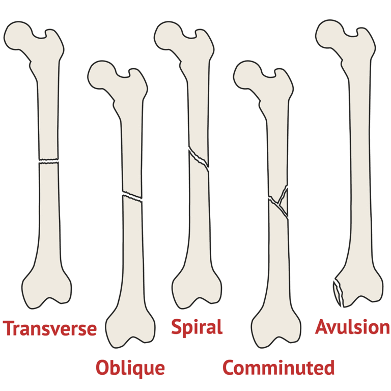
Avulsion: A fragment of bone is pulled away by the force of a tendon or ligament.
Potts Fracture: A specific fracture involving the bones of the ankle.
Colles fracture (distal radius fracture): A common fracture at the end of the radius bone in the forearm, near the wrist.
3. By Pattern
Transverse: The fracture line runs straight across the bone, perpendicular to its length.
Oblique: The fracture line runs at an angle across the bone.
Spiral: The fracture line wraps around the bone, often due to a twisting injury.
4. Miscellaneous
Green stick fracture: Common in children, where the bone bends and cracks but doesn’t break completely. One side of the bone is broken, while the other side is bent.
Depressed fracture: Bone fragments are pushed inward, below the level of the surrounding bone. This is often seen in skull fractures.
Comminuted fracture: The bone is broken into multiple small fragments. This usually occurs from high-impact trauma.
Displaced/overriding fracture: The broken ends of the bone are separated and overlap.
Impacted: One fragment of the fractured bone is driven into the other fragment.
Complicated fracture: The fracture is associated with significant damage to surrounding structures like nerves, blood vessels, joints, or muscles.
Stress fracture: A small crack in the bone that develops from repeated stress or overuse.
Pathological fracture: A fracture caused by an underlying disease that weakens the bone.
Signs and symptoms of fractures
Pain at the site of injury: Continuous pain that intensifies until the fractured bone is stabilized.
Local tenderness: Pain specifically when touching the area of the fracture.
Deformity: An abnormal shape or angulation of the injured limb due to the displacement or rotation of bone fragments. This can be felt or seen by comparing it to the uninjured limb.
Soft tissue swelling: Inflammation in the tissues around the fracture site.
Loss of function: Inability to use the injured limb properly because the muscles cannot work effectively without the support of intact bones.
Bruising: Discoloration of the skin due to bleeding under the surface.
Involuntary muscle spasms: Muscles around the fracture involuntarily contract.
Intense pain: Sharp pain, for instance, in the ribs when breathing deeply or coughing, indicating a possible fracture.
Abnormal mobility: Movement where there shouldn’t be movement, indicating a break.
Crepitus: A grating sound or sensation felt when the broken bone ends rub together.
Bone may be visibly seen protruding through the skin: In open fractures, the bone may be visible.
Impaired sensation/numbness: Loss of feeling due to nerve damage.
Shock: A serious condition that can occur due to significant blood loss.
Swelling and discoloration (Ecchymosis): Localized swelling and bruising as a result of trauma and bleeding into the tissues.
Assessment and Diagnostic Findings
The following methods are used to diagnose fractures:
History taking: Gathering information about how the injury occurred.
Physical Examinations: A thorough evaluation of the injured area.
X-ray examinations: Standard imaging technique to determine the location and extent of the fracture.
Bone scans, computed tomography (CT)/magnetic resonance imaging (MRI) scans: Advanced imaging techniques to visualize fractures in more detail, as well as any associated bleeding or soft-tissue damage.

Process of Fracture Healing
This process varies depending on the specific bone involved, the type of fracture, and the degree of movement at the fracture site. When rigid fixation isn’t used for tubular bones, healing typically progresses through five stages:
1. Tissue destruction & haematoma formation:
Following tissue damage, torn blood vessels lead to the formation of a haematoma. This is a collection of clotted blood located between the broken ends of the bones and within the surrounding soft tissues. A fibrin clot forms from fibrin, red blood cells, debris, and inflammatory fluids.
2. Inflammation & cellular proliferation:
This stage begins with acute inflammation and the buildup of inflammatory fluid. This fluid contains macrophages, which are cells that engulf and remove the haematoma and small pieces of bone that no longer have a blood supply. This phase lasts approximately five days. Fibroblasts, cells that produce connective tissue, move to the injury site, and granulation tissue (new connective tissue with tiny blood vessels) and new capillaries develop.
3. Stage of Callus formation (Soft Callus)
New bone begins to form as numerous osteoblasts (bone-forming cells) secrete spongy bone, which bridges the gap between the broken ends. Osteoclasts (bone-resorbing cells) start to remove any dead bone. This new deposit of bone and cartilage is called a callus. As this immature bone (soft callus) becomes more mineralized, the amount of movement at the fracture site gradually decreases.
4. Stage of consolidation (Hard callus)
Over the subsequent days, the callus matures. The cartilage within the callus is slowly replaced by new, stronger bone.
5. Stage of remodelling
This involves the reshaping of the callus through a continuous cycle of bone resorption (removal) and bone deposition (laying down new bone). The internal callus is remodeled to create a hollow marrow cavity, while the external callus is gradually broken down and removed. The bone continues to reshape, and eventually, the medullary canal (the central cavity of the bone) is reopened through the callus. The callus tissue is completely replaced with mature, dense compact bone. Often, the bone is thicker and stronger at the repair site than it was originally, making a second fracture more likely to occur at a different location.
Healing of Fractures
Factors necessary for bone healing
Haematoma formation: The initial blood clot is crucial for initiating the healing process.
Contact of bone ends and no interposition of other tissues: The broken ends need to be in close proximity without soft tissues or other materials blocking the gap.
Continued immobilization until callus is able to withstand stress: The fracture site needs to be kept still until the new bone is strong enough to handle normal forces.
Good supply of blood: Adequate blood flow is essential for delivering nutrients and cells needed for repair.
Enough rest of the fractured site: Allowing the injured area to rest promotes the healing process.
Good nutrition – calcium, proteins: Sufficient intake of calcium and protein provides the building blocks for new bone tissue.
Factors influencing bone healing.
(a). Systemic factors
Age: Healing is significantly faster in children compared to adults (almost twice as fast).
Activity level: Immobilization is necessary for proper healing.
Nutritional status: Adequate nutrition supports the body’s repair mechanisms.
Hormonal factors: Growth hormone (GH) promotes bone growth, while corticosteroids can inhibit it.
Diseases e.g. DM, anaemia, neuropathies: Conditions like diabetes mellitus, anemia, and nerve damage can impair healing.
Vitamin deficiencies e.g. A C D K: Vitamins A, C, D, and K are essential for bone health and repair.
Drugs e.g. anti coagulants, anti inflammatory: Medications like anticoagulants (blood thinners) and anti-inflammatory drugs can affect bone healing.
(b). Local factors.
Type of bone: Cancellous bone (spongy bone) heals more quickly than cortical bone (dense outer bone).
Type of fracture: Spiral fractures tend to heal better than transverse fractures.
Blood supply: Poor blood circulation to the fracture site hinders healing.
Reduction: Fractures heal faster when the broken bone ends are perfectly aligned (perfect reduction).
Infection: Infection at the fracture site significantly delays healing.
Soft tissue interposition: Soft tissues located between the broken bone ends can prevent proper union.
Mobilization: Both early and excessive mobilization can disrupt healing.
Management
Aims of management.
To regain and maintain the normal alignment of the injured part: Restoring the bone fragments to their correct anatomical position.
To regain normal function of the injured part: Restoring the ability to use the injured limb or area as before the injury.
To achieve the above objectives for the patient in the shortest time possible: Promoting efficient and timely healing.
Basic principles of managing fractures
The core principles of fracture management are:
Reduction
Immobilization
Rehabilitation
(1) Reduction: Reduction is the process of restoring the broken bone ends (and any fractured pieces) to their normal anatomical positions. This can be done through either open or closed manipulation of the injured area.
Closed reduction: This involves manually aligning the bone ends through manipulation and traction without surgery. X-rays are used to confirm the correct positioning. A cast is typically applied to keep the extremity immobilized and maintain the reduction.
Open reduction: This requires a surgical incision to directly visualize and align the bone fragments. Internal fixation devices are often used to hold the bones in the correct position while they heal.
(2) Immobilization: Immobilization is crucial for maintaining the reduction until the fracture has healed sufficiently. This can be achieved through external or internal fixation methods.
Methods of external fixation: These include casts, splints, and traction applied externally.
Internal fixation devices: These are devices surgically implanted to stabilize the fracture, such as pins, wires, screws, rods, nails, and plates.
(3) Rehabilitation: Rehabilitation focuses on regaining strength and restoring normal function to the affected area. The specific rehabilitation plan is tailored to the type of fracture and the reduction and immobilization methods used.
First aid management
This initial care is vital and can be life-saving. Following the ABCDs of life support is crucial:
Ensure that the patient is breathing, then proceed with:
Airway: Ensure the airway is clear.
Breathing: Ensure breathing is maintained.
Circulation: Check the pulse and control any bleeding.
Disability/Deformity: Immobilize any obvious deformities. Use splints to stabilize the limb, preventing further injury and pain.
Emergency Management of Fractures
Immediately after an injury where a fracture is suspected, it is crucial to immobilize the injured body part before moving the patient.
If an injured patient needs to be moved from a vehicle before splints can be applied, support the extremity both above and below the suspected fracture site to prevent rotation and angular movement.
Adequate splinting, including the joints above and below the fracture, is essential.
Movement of the fracture fragments causes more pain, further soft tissue damage, and bleeding.
Use temporary, well-padded splints that are firmly bandaged over the patient’s clothing to immobilize the fracture.
For lower extremity fractures, the legs can be bandaged together, using the uninjured leg as a splint for the injured one.
For upper extremity injuries, the arm can be bandaged to the chest, or an injured forearm can be placed in a sling.
Assess the neurovascular status distal to the injury to check the blood supply and nerve function in the periphery.
For open fractures, cover the wound with a clean (preferably sterile) dressing to prevent contamination of deeper tissues.
Do not attempt to realign the fracture, even if a bone fragment is protruding through the wound. Apply splints for immobilization.
In the emergency department, the patient will undergo a complete evaluation.
Clothing should be removed gently, starting with the uninjured side of the body and then the injured side. Cutting the clothing away may be necessary. Minimize movement of the fractured extremity to prevent further damage.
Management in hospital
Care depends on the classification and type of fracture.
Immobilization, reduction, and rehabilitation are performed.
Pain relief is provided.
Antibiotics are administered as needed.
Supportive treatments such as iron supplements (feso4), folic acid (FA), and multivitamins may be given.
Bone X-rays are performed.
Fluid resuscitation is initiated if necessary.
Infection prevention measures are implemented.
Nutrition, particularly calcium intake, is addressed.
Exercises and physiotherapy are prescribed.
Nursing care
Nursing Care
Encourage patients with closed (simple) fractures to resume their normal activities as soon as it is safe.
Teach patients how to manage swelling and pain related to the fracture and any soft tissue trauma. Encourage activity within the limitations imposed by the immobilization.
It is important to teach exercises to maintain the strength of unaffected muscles and to strengthen muscles needed for transfers and for using assistive devices (e.g., crutches, walker).
Teach patients how to use assistive devices safely. Plans are made to help patients modify their home environment if needed and to secure personal assistance if required.
Patient education includes self-care instructions, medication information, how to monitor for potential complications, and the need for ongoing healthcare follow-up.
Fracture healing and the restoration of full strength and mobility can take several months.
Managing Fractures at Specific Sites
The primary goal of management is maximum functional recovery.
Clavicle
Clavicle (collar bone) fractures are common injuries resulting from falls or direct blows to the shoulder.
Monitor the circulation and nerve function of the affected arm, comparing it to the unaffected arm to identify any neurovascular issues.
Advise the patient not to raise their arm above shoulder level until the fracture has healed (approximately 6 weeks).
Encourage elbow, wrist, and finger exercises as soon as possible and, when prescribed, shoulder exercises.
Inform the patient that vigorous activity should be limited for 3 months.
Humeral Neck
Humeral neck fractures are frequently seen in older women after falling on an outstretched arm. Perform a neurovascular assessment of the affected extremity to check for nerve and blood vessel involvement.
Teach the patient to support and immobilize the arm using a sling and swathe, which secures the supported arm to the trunk.
Start pendulum exercises as soon as the patient can tolerate them. Instruct the patient to avoid strenuous activity for an additional 10 to 14 weeks.
Inform the patient that some stiffness, aching, and limited range of motion may persist for 6 months or longer.
If a humeral neck fracture is displaced and requires fixation, exercises will begin only after a prescribed period of immobilization.
Humeral shaft fractures
Nerves and the brachial blood vessels can be injured, so neurovascular assessments are crucial.
Use well-padded splints for initial immobilization of the upper arm and support the arm at a 90-degree angle of elbow flexion. Use a sling or collar and cuff to support the forearm, and external fixators may be used for open fractures of the humeral shaft.
Functional bracing may also be used.
Teach the patient pendulum shoulder exercises and isometric exercises as prescribed.
Elbow
Elbow fractures (distal humerus) can injure the median, radial, or ulnar nerves.
Assess the patient for paresthesia (abnormal sensations) and signs of compromised circulation in the forearm and hand.
Closely monitor for Volkmann’s ischemic contracture (an acute compartment syndrome) and hemarthrosis (blood in the joint).
Reinforce information about the reduction and fixation of the fracture and the planned active motion once swelling subsides and healing begins.
Explain care if the arm is immobilized in a cast or posterior splint with a sling. Encourage active finger exercises.
Teach and encourage gentle range of motion exercises of the injured joint about 1 week after internal fixation.
Radial head fractures
These usually occur from a fall on an outstretched hand with the elbow extended.
Instruct the patient on how to use a splint for immobilization.
If the fracture is displaced, emphasize the need for postoperative immobilization of the arm in a posterior plaster splint and sling.
Encourage the patient to follow a program of active motion of the elbow and forearm as prescribed.
Wrist
Wrist fractures (distal radius [Colles’ fracture]) typically result from a fall on an open, dorsiflexed hand. They are common in elderly women with osteoporosis.
Reinforce cast care, or if wires were inserted for more severe fractures, teach incision care.
Instruct the patient to keep the wrist and forearm elevated for 48 hours after reduction.
Start active finger and shoulder motion promptly by teaching the following exercises to reduce swelling and prevent stiffness:
Hold the hand at heart level. Move the fingers from full extension to flexion. Hold and release. Repeat at least 10 times every hour while awake.
Use the hand in functional activities.
Actively exercise the shoulder and elbow through their full range of motion.
Assess median nerve sensory function by lightly pricking the distal aspect of the index finger and motor function by testing the patient’s ability to touch their thumb to their little finger.
If diminished circulation and nerve function are noted, address it immediately.
Hand and Fingers
Hand trauma often requires extensive reconstructive surgery. The goal is always to regain maximum hand function.
For non-displaced fractures, the finger is splinted for 3 to 4 weeks. Displaced and open fractures may require open reduction with internal fixation using wires or pins.
Encourage using the unaffected parts of the hand for function.
Evaluate the neurovascular status of the injured hand.
Teach the patient to control swelling by elevating the hand.
Pelvis
Pelvic fractures can result from falls, car accidents, or crush injuries. Most patients with pelvic fractures have other significant injuries.
Monitor for symptoms including bruising (ecchymosis), tenderness over the pubic symphysis, anterior iliac spines, iliac crest, sacrum, or coccyx, local swelling, numbness or tingling in the groin area and upper thighs, and inability to bear weight without pain.
Complete a neurovascular assessment of the lower extremities to detect injury to pelvic blood vessels and nerves.
As pain decreases, instruct the patient to gradually resume activity, using assistive devices for weight-bearing. Unstable pelvic fractures may require external fixation or open reduction and internal fixation (ORIF).
Focus on maintaining stable blood flow and comfort, and encourage early movement.
Examine urine for blood to assess for urinary tract injury. In male patients, do not insert a catheter until the urethra is confirmed to be intact.
Monitor for diffuse abdominal pain, hyperactive or absent bowel sounds, and abdominal rigidity or signs of free air or blood in the abdomen, which could indicate injury to the intestines or abdominal bleeding.
Monitor for hemorrhage and shock. Palpate both lower extremities for peripheral pulses, which may indicate a torn iliac artery or one of its branches.
Assess for injuries to the bladder, rectum, intestines, other abdominal organs, and pelvic vessels and nerves.
For stable pelvic fractures, maintain bed rest for a few days and manage symptoms until pain is controlled.
Provide fluids, dietary fiber, ankle and leg exercises, antiembolism stockings, logrolling techniques, deep breathing exercises, and skin care to prevent complications and increase comfort.
Monitor bowel sounds. If a patient has a coccyx fracture and experiences pain when sitting or during bowel movements, assist with sitz baths and administer stool softeners.
Femur and Hip
Femoral shaft fractures are most common in young adults involved in car accidents or falls from heights.
These patients often have multiple injuries and can develop shock from significant blood loss.
Assess the neurovascular status of the extremity, particularly blood flow to the lower leg and foot (check pulses and capillary refill). Doppler ultrasound may be used.
Note any signs of hip or knee dislocation and knee swelling, which may suggest ligament damage and knee instability.
Apply and maintain skeletal traction or a splint to relax muscles and align the fracture fragments before ORIF and later a cast brace.
Assist with partial weight-bearing as indicated, progressing to full weight-bearing as tolerated.
Reinforce that the cast brace should be worn for 12 to 14 weeks.
Instruct and encourage regular exercises for the lower leg, foot, and toes.
Assist with active and passive knee exercises as soon as possible, depending on the management approach and the stability of the fracture and knee ligaments.
Tibia and Fibula
Tibia and fibula fractures are the most common fractures below the knee, typically resulting from direct blows, falls with the foot flexed, or twisting motions.
Provide instructions on caring for a long leg walking cast or patellar-tendon-bearing cast.
Instruct and assist with partial weight-bearing, usually starting in 7 to 10 days.
Instruct on caring for a short leg cast or brace (used in 3 to 4 weeks), which allows knee motion.
Instruct on the care of skeletal traction if used.
Encourage hip, foot, and knee exercises within the limitations of the immobilization device.
Instruct the patient when to begin weight-bearing (usually around 4 to 8 weeks).
Instruct the patient to elevate the extremity to control swelling.
Perform ongoing neurovascular evaluations.
Rib
Rib fractures are frequent in adults and usually don’t impair function but cause painful breathing.
Assist the patient to cough and take deep breaths by supporting the chest with their hands or a pillow during coughing.
Reassure the patient that the pain will lessen significantly in 3 to 4 days and the fracture will heal within 6 weeks.
Monitor for complications such as atelectasis (collapsed lung), pneumonia, flail chest, pneumothorax, and hemothorax.
COMPLICATIONS OF FRACTURES
Early complications include;
Shock
Fat embolism
Compartment syndrome
Venous thromboembolism (deep vein thrombosis [DVT], Pulmonary embolism [PE])
Delayed Complications;
Delayed union
Malunion
Nonunion
Avascular necrosis (AVN) of bone
Reaction to internal fixation devices
Complex regional pain syndrome (CRPS, formerly called reflex sympathetic dystrophy (RSD))
Heterotopic ossification
MANIFESTATION OF COMPLICATIONS
Fat embolism syndrome:
Sudden onset, usually within 12 to 48 hours but can occur up to 10 days after injury.
Signs & symptoms: hypoxia, tachypnea, tachycardia, pyrexia; dyspnea, crackles, wheezes, precordial chest pain, cough, large amounts of thick white sputum.
Compartment syndrome:
Deep, throbbing, unrelenting pain not controlled by opioids.
Signs & symptoms include; cyanotic nail beds, pale or dusky and cold fingers or toes, prolonged capillary refill (greater than 3 seconds), diminished or absent pulse, motor weakness, paralysis, and paresthesia.
DIC – disseminated intravascular coagulation:
Unexpected bleeding after surgery.
Bleeding from mucous membranes, venipuncture sites, and the gastrointestinal and urinary tracts.
Infection: symptoms include:
Tenderness on examination
Pain
Redness
Swelling
Local warmth
Elevated temperature
Purulent drainage
Nonunion is manifested by:
Persistent discomfort and abnormal movement at the fracture site.
Risk factors include infection, tissue interposition, inadequate immobilization, or manipulation.
MANAGEMENT OF COMPLICATIONS
Treatment of shock:
Stabilize the fracture.
Restore blood volume and circulation.
Relieve pain.
Provide proper immobilization.
Protect from further injury.
Prevention and management of fat embolism:
Immediate immobilization of fractures.
Adequate support during turning and positioning.
Maintenance of fluid and electrolyte balance.
Prompt respiratory support.
Corticosteroids and vasopressor medications may be given.
Compartment syndrome:
Control swelling by elevating the extremity to heart level or releasing restrictive devices.
A fasciotomy may be needed.
The wound remains open and covered with moist sterile saline dressings for 3 to 5 days.
The limb is splinted and elevated.
Prescribed passive range-of-motion exercises may be performed every 4 to 6 hours.
Nonunion:
Treated with internal fixation, bone grafting, electrical bone stimulation, or a combination.
Related Question
Josephine a thirty-year-old female patient has been involved in a road traffic accident and sustained a compound fracture.
Outline ten signs and symptoms of fracture.
Discuss the negative factors that can influence the healing of a bone.
Describe the healing of a bone.
Mention ten complications of fractures.
SOLUTIONS
a)
Pain aggravated by movement
Tenderness over the fractured limb
Loss of function of the affected part or the whole limb
Deformity
Shortening of the limb
Abnormal mobility at the affected area
Crepitus or grating of the bone ends
Swelling of the affected part
Shock may occur
The bone may be visible (in a compound fracture)
b)
Tissue fragments between bone ends.
Deficient blood supply.
Poor alignment of bone ends.
Continued mobility of bone ends.
Infection.
Systemic illness.
Malnutrition.
Drugs e.g. Corticosteroids.
Aging.
c)
Hematoma forms.
Inflammation and cellular proliferation occur.
Soft callus forms.
Hard callus forms.
Remodeling occurs.
d)
Hemorrhage/shock.
Fat embolism.
Infections.
Hypostatic Pneumonia.
Damage to nearby structures.
Keloids.
Loss of function.
Damage to the nerves.
Necrosis.
Delayed union/Malunion/Nonunion.
We are a supportive platform dedicated to empowering student nurses and midwives through quality educational resources, career guidance, and a vibrant community. Join us to connect, learn, and grow in your healthcare journey
Quick Links
Our Courses
Legal / Policies
Get in Touch
(+256) 790 036 252
(+256) 748 324 644
Info@nursesonlinediscussion.com
Kampala ,Uganda
© 2026 Nurses online discussion. All Rights Reserved