Bone Conditions in Children
Subtopic:
Osteopenia of Prematurely

Osteopenia of prematurity is a bone-weakening condition in premature infants. It occurs due to reduced calcium and phosphorus levels in the bones, making them fragile and prone to fractures.
Premature birth significantly disrupts the normal process of bone mineralization and growth, leading to this condition.
Normal bone formation involves the deposition of minerals, mainly calcium (Ca<sup>+2</sup>) and phosphorus (P), onto an organic framework (osteoid) produced by osteoblasts (the cells responsible for building bone). Osteoclasts (the cells involved in breaking down bone) are also crucial for bone remodeling.
Osteopenia in premature infants primarily arises from an inadequate intake of calcium to support the rapid bone growth that should be occurring in the womb.
Causes
Insufficient vitamin D in the mother during pregnancy can contribute. Vitamin D is vital for calcium absorption in the intestines and kidneys, and it’s transferred from mother to fetus.
Premature infants experience increased phosphorus loss in their urine compared to full-term infants.
Liver conditions can lead to vitamin D deficiency. For example, cholestasis, which involves a blockage of bile flow, can impair vitamin D absorption.
Signs and Symptoms
Clinically, osteopenia often becomes noticeable between 6 and 12 weeks of age. It can be asymptomatic, but severe cases may present with:
Severe manifestations
Poor weight gain and a failure to grow at the expected rate.
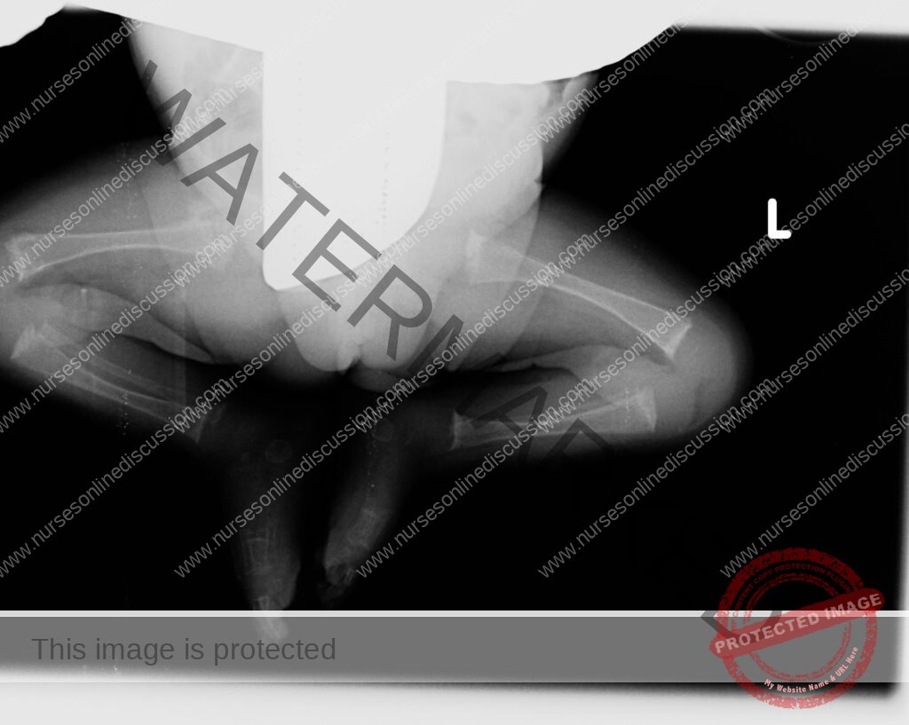
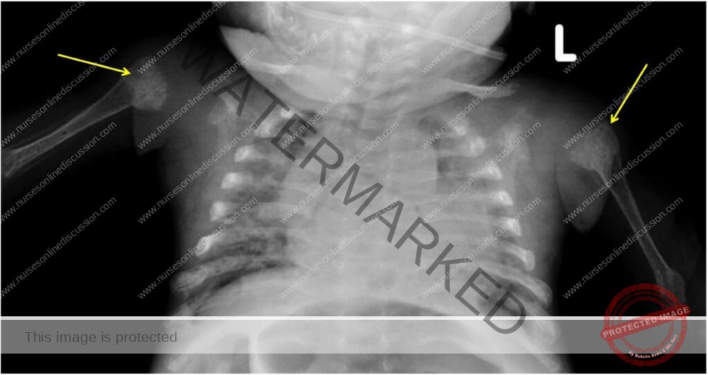
Signs resembling rickets, such as slowed growth, a prominent forehead (frontal bossing), and widening of the ends of long bones (epiphyseal widening).
Fractures may be suspected if the infant shows pain upon handling.
Breathing difficulties or an inability to come off ventilator support can occur due to a poorly compliant chest wall.
Consequences of osteopenia. This condition can potentially lead to nearsightedness (myopia) in premature infants due to skull shape changes. Later in childhood, affected infants may remain thinner and shorter, with lower bone mineral content (BMC) and density. Increased calcium excretion in urine has also been observed.
Decreased spontaneous movements.
Swelling of the arms or legs might indicate fractures that haven’t been diagnosed.
Pathophysiology
Bone mineralization starts during the embryonic stage, but the majority of this process happens during the third trimester of pregnancy.
Most of the calcium and phosphorus transfer from mother to fetus occurs in the third trimester, particularly around the 34th week of gestation, when approximately 80% of the mineral content is accumulated.
By the 28th week of gestation, mineral accumulation is around 60mg per day, increasing significantly to over 300mg per day between the 35th and 38th weeks.
Osteoblasts produce the organic bone matrix for calcium and phosphate deposition, leading to bone growth through increased trabecular thickness. This process is essential for the baby’s development. If this mineral deposition is insufficient, it results in osteopenia of prematurity, potentially causing fractures in long bones, including the ribs, which can lead to breathing problems.
A premature infant may not receive adequate calcium and phosphorus needed for strong bone development.
Premature babies excrete more phosphorus in their urine, and their limited movement further contributes to weakened bones.
Risk Factors
Fetal and neonatal causes
Prematurity and birth weight: Early birth leads to calcium and phosphorus deficits. The likelihood of osteopenia increases as gestational age and birth weight decrease. These infants have higher nutritional and growth demands coupled with mineral deficiencies.
Feeding practices: Delayed initiation of feeding through the digestive tract (enteral feeding), prolonged reliance on intravenous nutrition (parenteral nutrition), using unfortified breast milk, restricting enteral feeding, and malabsorption issues can all cause mineral deficiencies.
Human milk: Breast milk is naturally low in phosphorus, and donor milk often has even lower levels than milk from the infant’s own mother. Extended use without supplementation can result in low phosphate levels in the blood, hindering incorporation into the bone structure. Unfortified breast milk cannot provide the mineral accumulation rates achieved through placental transfer.
Drugs: Medications like corticosteroids, furosemide, and methylxanthines, commonly used in preterm infants, can cause calcium to be released from the bones, reducing bone mineral content.
Lack of mechanical stimulation: Bone growth is stimulated by movement and weight-bearing. Premature birth, illness, sedation, and paralysis disrupt this stimulation. Infants with neurological impairments like spina bifida, who have limited mobility, are also at risk of poor bone growth.
Vitamin D: Vitamin D deficiency after birth can occur in breastfed infants not receiving supplements due to low levels in breast milk (25–50 IU/L). Other reasons for vitamin D deficiency include:
Renal (kidney) disorders causing osteodystrophy (abnormal bone development).
Drugs like phenytoin and phenobarbital, which increase vitamin D breakdown.
Aluminum contamination: Aluminum present in parenteral nutrition solutions can interfere with bone mineralization.
Malabsorption: Conditions like prolonged cholestasis and short gut syndrome can impair the absorption of vitamin D and calcium.
B. Maternal factors
Low vitamin D levels in the mother result in lower levels in the fetus.
Maternal smoking, being underweight, low calcium intake, and increased physical activity during the third trimester can lead to decreased calcium in the fetus.
Exposure to high doses of magnesium in the womb, preeclampsia (high blood pressure during pregnancy), chorioamnionitis (infection of the amniotic sac), and placental infections are linked to osteopenia.
Infants with intrauterine growth restriction (IUGR), where the placenta may be damaged, are more likely to develop postnatal rickets due to altered phosphate transport.
Having multiple pregnancies and having a male infant are associated with a higher incidence.
Placental hormones like estrogen and parathyroid hormone (PTH), and PTH-related protein also play a role in fetal bone development.
Predisposing factors
Premature birth, especially before 30 weeks of gestation.
Prolonged use of medications that increase mineral excretion, such as diuretics and theophylline.
Vitamin D deficiency.
Low levels of parathyroid hormone during pregnancy, which can suppress fetal calcium and phosphorus levels.
Inefficient calcium and phosphorus intake in infants with poor tolerance to enteral feeds who require total parenteral nutrition.
Common newborn illnesses like sepsis, acidosis, and necrotizing enterocolitis can disrupt bone remodeling by reducing osteoclast activity, decreasing calcium absorption, and increasing calcium loss through the kidneys.
Paralysis may increase calcium loss through the kidneys.
Short gut syndrome, leading to malabsorption of vitamin D and calcium.
Diagnosis and Investigations
Radiographs (X-rays): Often the initial way osteopenia is identified, though assessment can be subjective.
Calcium levels: May remain within the normal range until the condition is quite advanced.
Phosphorus: Blood phosphate levels are typically low (below 3 mg/dL).
Ultrasound: This offers advantages like easy access and no radiation exposure. It’s used on peripheral sites like the heel bone (calcaneus) and shin bone (tibia). It can assess both the quality and quantity of bone, including mineralization and cortical thickness, as well as bone mass, elasticity, and internal structure.
Dual-energy x-ray absorptiometry (DEXA): يعتبر المعيار الذهبي لتقييم حجم العظام وحالة المعادن فيها ويمكن أن يتنبأ بخطر الكسور عند الأطفال حديثي الولادة. ومع ذلك، فإن القيود في استخدامه وتفسير البيانات تمنع التطبيق السريري الواسع النطاق. (DEXA is the gold standard for assessing bone size and mineral status and can predict fracture risk in newborns. However, limitations in its use and data interpretation hinder widespread clinical application.)
Management
Aims
To restore normal calcium and phosphorus levels in the body.
To prevent further complications or worsening of the condition.
Admission
The infant is admitted to the pediatric ward if referred from outside the hospital.
Assessment
Demographic information is collected, including the baby’s name, age, sex, etc.
A detailed medical and pregnancy history is taken, including prenatal and birth details, birth weight, and Apgar scores at birth.
A thorough physical examination is performed, focusing on bone formation to identify any abnormalities.
Immediate care
The baby is placed in a comfortable, warm bed to prevent hypothermia.
Pain relievers like paracetamol (2.5mg every 8 hours for three days) are given to manage potential pain from undiagnosed fractures.
If fractures are suspected, the affected limb is immobilized to maintain bone alignment. The doctor is notified for assessment and to order necessary investigations for diagnosis.
Investigations will include:
Blood tests to measure calcium, phosphorus, and a protein called alkaline phosphatase.
Ultrasound to check for fractures.
X-rays to determine the extent of any fractures.
TREATMENT
The following treatments are typically administered as prescribed by the doctor:
Calcium (1.25mmol/kg per dose) added to intravenous fluids like normal saline or Ringer’s lactate until the condition stabilizes.
Intravenous phosphorus (1mmol/kg per dose) added to intravenous fluids until the condition stabilizes.
Vitamin D supplements are provided, especially for infants with liver problems.
Nursing interventions
Ensure the baby is kept warm and comfortable. Monitor vital signs (temperature, pulse, respiration rate).
Ensure the baby receives a diet rich in calcium and phosphorus, often through fortified milk.
Encourage physical exercises as recommended by physiotherapists.
Provide a quiet and comfortable environment to ensure adequate rest and sleep.
Provide psychological support to the mother to reduce anxiety.
Promote both environmental and personal hygiene to prevent infections.
Administer medications as prescribed by the doctor. Monitor urine calcium and phosphorus levels weekly. Discharge is considered when the patient’s condition improves.
Advise on discharge
Advise the mother to continue feeding the baby a diet rich in calcium and phosphorus. Emphasize the need to handle the baby gently due to the risk of fractures.
Schedule a follow-up appointment to monitor the baby’s progress and the prognosis of the condition.
We are a supportive platform dedicated to empowering student nurses and midwives through quality educational resources, career guidance, and a vibrant community. Join us to connect, learn, and grow in your healthcare journey
Quick Links
Our Courses
Legal / Policies
Get in Touch
(+256) 790 036 252
(+256) 748 324 644
Info@nursesonlinediscussion.com
Kampala ,Uganda
© 2026 Nurses online discussion. All Rights Reserved