Reproductive Health
Subtopic:
Examination of placenta
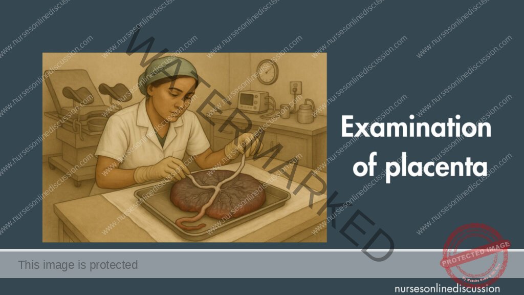
A thorough examination of the placenta and fetal membranes (amnion and chorion) immediately after their delivery (third stage of labor) is a crucial component of postnatal care. This routine procedure provides valuable information about the completed pregnancy, potential risks for the mother or newborn postpartum, and can sometimes offer insights into fetal well-being or pregnancy complications.
Purpose of Placental Examination:
Ensure Completeness:
The primary goal is to confirm that the entire placenta and all membranes have been expelled. Retained placental fragments or membranes are a significant cause of postpartum hemorrhage (both immediate and delayed) and postpartum infection (endometritis).
Identify Abnormalities:
To detect any structural abnormalities of the placenta, umbilical cord, or membranes that might have implications for maternal or neonatal health.
Some abnormalities may suggest underlying conditions or risks that require further investigation or follow-up.
Provide Clinical Information:
The placenta can provide clues about intrauterine events, such as infection, infarction (tissue death due to lack of blood supply), or issues with fetal growth.
Medico-Legal Documentation:
A record of the placental examination is part of the comprehensive birth record.
Guide Postpartum Management:
Findings can influence decisions about postpartum maternal care (e.g., need for uterine exploration if incompleteness is suspected) or neonatal observation/investigation.
Procedure for Placental Examination:
The examination should be performed systematically in a well-lit area, usually on a clean, flat surface. Standard precautions (gloves) should be observed.
I. Examination of the Umbilical Cord:
Number of Vessels:
Carefully inspect the cut end of the umbilical cord.
Normal: Two umbilical arteries and one umbilical vein (AVA). The arteries are usually smaller with thicker walls, while the vein is larger with a thinner wall and may remain open.
Abnormal: A single umbilical artery (SUA) or two-vessel cord is the most common abnormality (occurring in about 1% of pregnancies). While many babies with SUA are healthy, it can be associated with an increased risk of congenital anomalies (especially renal, cardiac, and chromosomal abnormalities) and intrauterine growth restriction (IUGR). Its presence should prompt careful neonatal assessment.
Insertion Site:
Observe where the umbilical cord inserts into the fetal surface of the placenta.
Normal (Central/Eccentric): Usually inserts near the center or slightly off-center.
Marginal Insertion (Battledore Placenta): Inserts at the very edge of the placenta (like a battledore racket). Generally not problematic but may be associated with a slightly increased risk of cord compression during labor or preterm birth.
Velamentous Insertion: The umbilical vessels run through the membranes (amnion and chorion) before inserting into the placenta. These vessels are unprotected by Wharton’s jelly and are vulnerable to rupture or compression, especially if they traverse the cervical os (vasa previa). Vasa previa is a serious obstetric emergency associated with high fetal mortality if the vessels rupture during labor.
Length:
Normal length is approximately 50-60 cm.
Short Cord (<35 cm): May be associated with fetal movement restriction, abruptio placentae, or uterine inversion.
Long Cord (>80-100 cm): May be associated with true knots, nuchal cords (cord around the neck), cord prolapse, or fetal entanglement.
Knots:
False Knots: Kinks or twists in the vessels that are not true knots and are of no clinical significance.
True Knots: Result from fetal movement through a loop of cord. Can tighten during labor, obstructing blood flow and potentially leading to fetal distress or demise, though many are loose and cause no problems.
Other Abnormalities:
Strictures or Torsion: Twisting or narrowing of the cord.
Hematomas or Cysts.
Wharton’s Jelly: Observe the amount (scant Wharton’s jelly can make the cord more prone to compression).
II. Examination of the Fetal Membranes (Amnion and Chorion):
Completeness:
Hold the placenta by the cord, allowing the membranes to hang down. Examine them for completeness. They should form a sac.
Identify the point of rupture (the hole through which the baby was delivered).
Carefully inspect for any ragged edges or missing pieces, which might indicate retained membranes in the uterus.
Color and Clarity:
Normal: Translucent, thin, and glistening.
Meconium Staining: Greenish or yellowish staining indicates the passage of meconium (fetal stool) in utero, which can be a sign of fetal distress or post-maturity. The duration of staining (e.g., on the fetal surface vs. deeply into the membranes) can give an idea of timing.
Cloudiness or Opacity with Foul Odor: Suggests chorioamnionitis (intrauterine infection).
Insertion:
Note how the membranes insert onto the placental disc (e.g., circumvallate placenta, where membranes insert inward from the edge).
III. Examination of the Placental Disc:
Fetal Surface (Facing the Baby):
Appearance: Normally smooth, shiny, and grayish-blue, covered by the amnion. Umbilical vessels radiate from the cord insertion site across this surface.
Abnormalities:
Amnion Nodosum: Small, opaque nodules on the amnion, associated with oligohydramnios (low amniotic fluid).
Subchorionic Fibrin Deposits: Yellowish-white deposits beneath the amnion, usually of no clinical significance unless extensive.
Cysts or Tumors: Rare.
Maternal Surface (Attached to the Uterine Wall):
Appearance: Normally deep red, rough, and divided into 15-20 lobes or cotyledons by grooves (sulci).
Completeness: This is a critical part of the examination. Carefully inspect all cotyledons to ensure they are present and intact. Check if any appear “shaggy” or if a piece is missing. A missing cotyledon or fragment necessitates further investigation (and possibly manual exploration of the uterus) as it indicates retained placental tissue.
Abnormalities:
Infarcts: Localized areas of dead tissue, appearing as firm, pale, or yellowish-white areas. Small, peripheral infarcts are common and usually benign. Large or central infarcts can impair placental function and may be associated with conditions like pre-eclampsia or IUGR.
Calcifications: Gritty, white deposits, more common in post-term pregnancies. Usually not clinically significant unless extensive.
Hematomas (Blood Clots):
Retroplacental Hematoma: Clot on the maternal surface, indicative of abruptio placentae (premature separation of the placenta). The size and age of the clot can be informative.
Accessory Lobes (Succenturiate Lobes): One or more smaller lobes of placental tissue separate from the main placental disc, connected by fetal vessels running through the membranes. These are important to identify because if an accessory lobe is retained in the uterus after delivery of the main placenta, it can cause PPH or infection. The membranes should be inspected for torn vessels leading to an empty space, suggesting a retained succenturiate lobe.
Placenta Accreta Spectrum: Abnormally deep attachment of the placenta to the uterine wall. This is a serious condition often diagnosed antenatally, but incomplete separation or inability to deliver the placenta may be the first sign. Examination may reveal no clear plane of separation on the maternal surface.
Shape and Size:
Shape: Typically round or oval. Abnormal shapes (e.g., bilobed, trilobed, battledore) are noted.
Dimensions: Measure diameter and thickness (typically 2-4 cm thick at the center).
Weight: Average weight is about 450-550 grams (or roughly 1/6th of the baby’s weight). A very small placenta may be associated with IUGR, while a large, edematous placenta can be seen with conditions like hydrops fetalis or maternal diabetes.
Documentation:
All findings from the placental examination should be documented accurately in the mother’s and/or baby’s medical record. This includes weight, dimensions, cord details (vessels, insertion, knots), membrane appearance, and completeness of cotyledons.
Any abnormalities found and any actions taken (e.g., suspicion of retained products) must be clearly recorded.
When to Send for Pathological Examination:
While routine pathological examination of all placentas is not standard practice in many settings, it is indicated in specific circumstances, such as:
Stillbirth or neonatal death
Preterm birth
Intrauterine growth restriction (IUGR)
Suspected intrauterine infection (chorioamnionitis)
Severe pre-eclampsia or eclampsia
Multiple gestations with complications
Maternal systemic illness (e.g., severe diabetes, autoimmune disease)
Neonatal neurological problems
Known or suspected fetal anomalies
Gross placental abnormalities noted on routine examination
Recurrent pregnancy loss
Related Topics
- Reproductive Health
- Pillars of Safe Motherhood
- Methods of Family Planning
- Management of STI’s/HIV/AIDS
- Adolescent Health and Development
- Adolescent and Reproductive Health
- Adolescent Friendly Health Services
- Post Abortion Care
- Signs and Symptoms of Pregnancy
- Signs and Symptoms of Labor
- Management of 2nd Stage of Labor
- Management of 3rd Stage of Labor
- Care of a Baby’s Cord
- Health Education of Mothers
- Referral System for Mother
- Signs and symptoms of 3rd stage of labor
- Examination of placenta
- Identification of mothers at risk and their referral
We are a supportive platform dedicated to empowering student nurses and midwives through quality educational resources, career guidance, and a vibrant community. Join us to connect, learn, and grow in your healthcare journey
Quick Links
Our Courses
Legal / Policies
Get in Touch
(+256) 790 036 252
(+256) 748 324 644
Info@nursesonlinediscussion.com
Kampala ,Uganda
© 2026 Nurses online discussion. All Rights Reserved