Communicable Diseases
Subtopic:
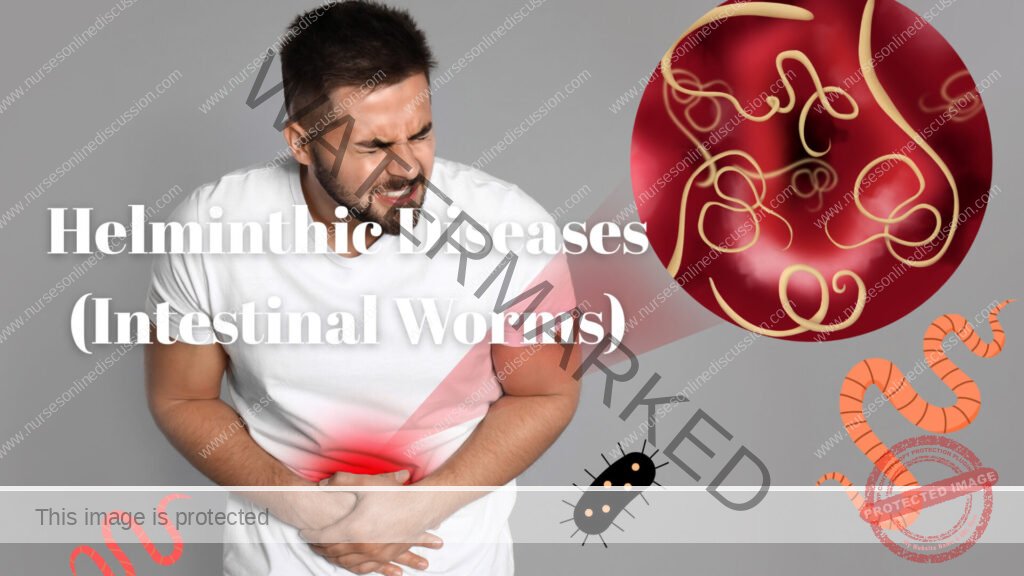
Helminthic Diseases (Intestinal Worms)

HELMINTHIC DISEASES (WORMS)
These are diseases caused by worms. There are three groups of worms and these are;
- Nematodes (round worms)
- Cestodes (tape worms)
- Trematodes (flukes)
All worms have human beings as their definitive host. In humans, adult worms reside in the intestines and they lay eggs which are passed in stool. Thus proper disposal of stool is one of the key preventive measures of worms.
NEMATODES
These worms are cylindrical and elongated. Examples of Nematodes are;
- Ascaris lumbricoides (round worm)
- Strongyloides starcolaris
- Ankylostoma duodenale
- Necator americanus hook worms
- Enterobius vermicularis (thread worms)
- Filarial worms
ASCARIASIS
It’s a common Nematode infection of the small intestines caused by Ascaris lumbricoides. This worm is the one commonly called round worm.
Epidemiology
The disease is common in Africa. It affects children more and more heavily than adults, because of their habit of putting whatever they come across in their mouth.
Mode of transmission
It’s basically faecal-oral route, by ingestion of food, fruits or drinking water that contains eggs.
Life cycle
- Large and adult round intestinal worms live in the small intestines of human beings, where they lay eggs. A female may lay up to 200,000 eggs a day. These eggs are then passed out in stool. To be infective, they have to get embryonated and this takes place in loose soil, which is not too dry in 8-50 days. Oxygen must be available in such soil.
- The embryonated eggs can be carried away from the contaminated place into houses by feet, foot wear or dust by wind. Human being can then swallow such eggs after contaminating food, water, fruits e.t.c. and after this, they hatch in the intestinal canal to yield larvae that penetrate the intestinal wall to access the blood stream.
- The larvae then follow the blood stream through the portal system to the liver and to the right side of the heart which sends them to the lungs via the pulmonary artery.
- Within the lungs, they penetrate the pulmonary capillaries to access the air ways i.e. alveoli, bronchioles, bronchi, trachea and then the pharynx. They get re swallowed to return to the GIT and then settle in the Jejunum, where they mature into adult worms in about 2 months.
Clinical features
Symptoms are always vague or absent
- Vague abdominal discomfort
- Adult worm can be passed in stool
Other temporary symptoms occur during passage of larvae through the lungs e.g.
- Cough
- Fever
- Wheezing
- Shortness of breath
- Allergic dermatitis
Complication
- Intestinal obstruction e. worms get tangled at a site in the small intestines to cause obstruction
- Obstructive jaundice 2° to obstruction of the bile ducts by the migrating larvae
- Liver abscess can be set up by larvae that remain hiding in the liver
- Malnutrition, because worms compete with human beings for the nutrients in the small intestines
Investigations
- Stool microscopy-shows eggs typical of Ascaris lumbricoides
- Full blood count-can show an eosinophilia
Management
- The drug of choice is Adult dose 500mg single dose or 100mg twice for 3/7
- Albendazole 400mg single dose
- An alternative is Levamisole (ketrax) 5mg/kg single dose
- Manage complications appropriately g. surgical measures for I.O, good nutrition for malnutrition
Prevention and control
Good environmental health
- Proper disposal of children’s faeces
- Training children to use latrines
- Washing of hands after toileting and before handling food
- Washing of fruits and vegetables before eating
- Having drying racks for utensils well above soil and dust
Routine deworming every 2-3 months is a better way of protection against worms
ANKYLOSTOMIASIS (HOOK WORM DISEASE)
Infection with hook worms varies from asymptomatic infection to chronic and disabling disease due to iron deficiency anemia. There are two types of hook worms and these are;
- Ankylostoma duodenale
- Necator americanus
Epidemiology
Hook worms are common in hot humid areas of Africa. They need a mean temperature of 18°c. The extent to which one develops anemia due to hook worms depends on nutritional status, daily iron intake and the total load of worms in one’s intestines
Mode of transmission
Via skin penetration by the larval forms of the worms
Life cycle
- Adult worms live in the intestines of human beings, attached there by using hooks on their heads. These worms lay eggs which are already embryonated as they pass in stool. Once outside, they develop into larvae called Rhabiti form larvae, which are not infective yet they feed highly while in soil.
- These larvae eventually transform to another form of larvae within 5 days, called Filari form larvae which are non- feeding.
- These larval forms attach themselves on to the grass or hide in the soil. However they have a tendency to attach themselves to whatever touches them. Therefore when a human being’s foot/leg touches them, they attach to it and actively penetrate through the skin to gain access to the venous system. They then follow the same tract as Ascaris lumbricoides (hepato-pulmonary tract, then mouth and back to the intestines) where they mature into adult worms
- The whole cycle can take up to 40 days. The adult worms attach to the intestinal mucosa by their hook-like teeth in their mouth.
Clinical picture
Most hook worm infections are asymptomatic. Patients also present with;
- Vague abdominal pain
- Abdominal distension
- Diarrhea mixed with blood
Other features are due to complications e.g.
- Severe pallor due to iron deficiency
- Oedema due to loss of proteins
- Tachycardia and murmurs
- Itching and erythematous papules at sites where larvae penetrate skin
Complications
- Severe The hook worms suck blood from the intestines (Ankylostoma duodenale sucks 0.3 ml/day and Necator americanus sucks 0.2ml/day)
- Cardiac failure secondary severe anemia
- Oedema due to loss of proteins leading to hypoproteinuria
- Gut perforation
- Intestinal obstruction
Investigations
- Stool microscopy-shows eggs typical of hook worms
- Full blood count-shows an eosinophilia and haemoglobin levels
Management
- Mebendazole and Albendazole are still the drugs of choice (Dose as in Ascaris)
- Correct anemia with iron replacement therapy of ferrous sulphate, Adults 200mg TDS for about a mouth
Folic acid can be added
- High protein diet to replace what is lost
- Blood transfusion should be avoided and should be taken as last resort with very low Hb
Prevention and control
- Wear shoes to prevent entry of larvae through the skin
- The rest of the measures are like for Ascariasis
ENTEROBIASIS
It’s an intestinal disease caused by Enterobius Vermicularis. It’s common in the tropics and more in school children because of their life styles
Mode of transmission
Faecal- oral route
Life cycle
- Initial infection follows ingestion of eggs of the adult worms (thread worm) in the contaminated food, drinks, fruits etc. However, infection can be maintained by direct transfer of the effective eggs from the anus to the mouth (auto infection)
- Air borne infection through inhalation of the eggs is possible but rare. After ingestion, eggs hatch in the stomach and small intestines. The resulting larvae mature into an adult worm in the lower intestines and the lower colon without going through the Hepato-pulmonary route.
- Gravid adult worms migrate to rectum to discharge the eggs on the peri-anal skin especially during the night. This causes anal itching and scratching leading to established auto infection. It takes 3-6 weeks.
Clinical features
Pruritus ani (anal itching) is the main symptom which can provoke anal scratching with consequent excoriation and secondary bacterial infection.
General symptoms
- sleep disturbance
- restlessness
- loss of appetite
- weight loss
Investigations
Apply an adhensive tape over the anus in the morning and stick the tape to a slide and observe under a microscope to look for the eggs characteristic of pin/thread worms.
Management
Drug of choice is still either Mebendazole or albendazole It’s better to treat the whole family
Prevention and control
- Good personal hygiene g.
- regular bathing and hand washing
- cut finger nails short
- wash under clothes like pants and knickers, as well as night clothes and bed clothes
- Avoid and correct overcrowding
- Practice proper disposal of faeces
- Treat the whole family once one person is suspected to be having the disease
- Give health education to the infected individuals on how to prevent or avoid re-
CESTODES
Also called tape worms. Generally they all have arounded head called scolex and a flat body of multiple segments called proglottids. The scolex is specialized to attach on to the intestinal wall by suckers and hooks.
These worms grow by adding new proglottids to the germinal end next to the scolex, while the oldest proglottids at the distal end are usually gravid and contain eggs and are the ones excreted in the faeces
There are two major tape worms
- Taenia solium (pig tape worm)
- Taenia saginatta (beef tape worm) These worms cause a disease called Taeniasis
TAENIASIS
It’s an infection of humans by tape worms. Almost all cases I the tropics are due to Taenia solium and Taenia saginatta
Disease is common in areas where pork and beef are eaten when still raw or poorly cooked. The worms have an intermediate host, which is a pig for Taenia solium and cattle for Taenia saginatta
Life cycle
- Adult worms live in the intestines of human beings. Broken segments (proglottids) containing a gravid uterus with or without eggs then fall off the adult worms and are passed in stool.
- These eggs or proglottids are then ingested by cattle (for Taenia saginatta) or pigs (for Taenia solium) and while in the GIT, the eggs hatch to yield embryos (larvae) that penetrate the bowel wall to gain access to the blood stream, and later they are carried to striated muscles of the respective animals in blood.
- Within the striated muscles, the larvae invaginate, grow and form infective cysts called
- cysticerci, within pork or beef and this meat is infective to humans
- When a human being ingests meat or pork which is raw or under cooked and contain the cysticerci, the cysts get dissolved by the gastric acid in the stomach to release the embryos.
- The Taenia saginatta embryo attaches itself on the wall of the small bowel by its head and grows into an adult worm.
- The Taenia solium embryos behave differently, penetrating the bowel wall to get carried in the blood stream to organs like striated muscles and brain and this sets up an extra intestinal disease called Cysticercosis due to Taenia solium
- Note; For Taenia solium, eggs can hatch on grass and the ingested larvae are still infective to pigs, unlike for Taenia saginatta where only eggs cause infection.
Clinical picture
Usually asymptomatic, but some people may have;
- loss of weight
- abdominal discomfort
- anal itching
Taenia solium infections are more dangerous, especially when the cysticerci invade the brain. Patient may present with;
- epilepsy
- muscular pains
- Neurological signs like hemiplegia, headache, and
Investigations
Stool microscopy- can show either the segments or the eggs specific of Taenia Management
Praziquantel is the drug of choice. It’s given as single dose of 5-10mg/kg Niclosamide is the alternative if Praziquantel is not available
Dose; Adult 2g (4tablets) single dose Children 11-34kg 2tablets single dose
Prevention and control
- proper disposal of faeces
- encourage people to avoid eating raw meat and pork and encourage proper cooking of such meat
- proper disposal of carcases in pits
- treat all individuals in a given catchment area suspected to be infected Taenia
- personal hygiene is of course important g. hand washing before eating, after eating and after toileting
Get in Touch
(+256) 790 036 252
(+256) 748 324 644
Info@nursesonlinediscussion.com
Kampala ,Uganda
© 2025 Nurses online discussion. All Rights Reserved Design & Developed by Opensigma.co

