Anatomy & Physiology 2
Subtopic:
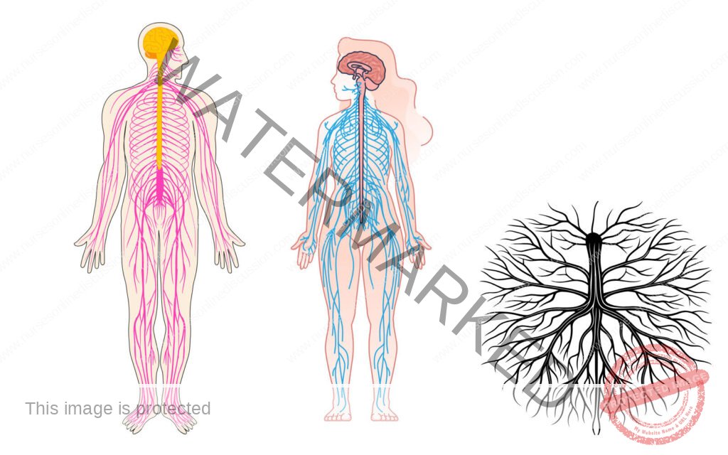
The Nervous System
The Nervous System
General Function
- None of the body system is capable of functioning alone. All are interdependent and work together as one unit so that normal conditions within the body may prevail.
- Control of the body’s billions of cells is accomplished mainly by two communication systems: the nervous system and the endocrine system.
- Both systems transmit information from one part of the body to another, but they do it in different ways. The nervous system transmits information very rapidly by nerve impulses conducted from one body area to another.
- The endocrine system transmits information more slowly by chemicals secreted by ductless glands into blood steam and circulated from glands to other parts of the body. The nervous system serves as the chief coordinating agency.
- Conditions both within and outside the body are constantly changing; the purpose of the nervous system is to respond to these internal and external changes (known as stimuli) and so cause the body to adapt to new conditions. It is through the nerve impulse sent to the various organs by the nervous system that a person’s internal harmony and the balance between the person and the environment are maintained.
- The nervous system has been compared to a telephone exchange, in that the brain and the spinal cord act as switching centers and the nerve trunks act as cables for carrying messages to and from these centers.
Cells of nervous system and their functions
The two types of cells found in the nervous system are called neurons or nerve cells and neuroglia, which are specialized connective tissue cells. Neurons conduct impulses, whereas
Neuroglia supports neurons.
Neurons
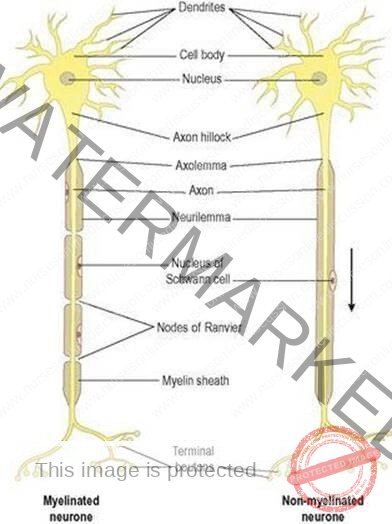
- Each neuron consists of three parts: a main part called the neuron cell body, one or more branching projections called dendrites, and one elongated projection known as an axon.
- Dendrites are the processes or projections that transmit impulses to the neuron cell bodies, and axons are the processes that transmit impulses away from the neuron cell bodies.
- Myelin sheath is a white, fatty substance formed by Schwann cells that wrap around some axons outside the central nervous system. Such fibers are called myelinated fibers.
- While Nodes of Ranvier are indentions between adjacent Schwann cells.
- Neurons can be classified structurally and functionally. The three types of functional classification of neurons are according to the direction in which they transmit impulses.
- These are: sensory neurons, motor neurons, and interneuron.
Sensory neurons transmit impulses to the spinal cord and brain from all parts of the body.
Motor neurons transmit impulses in the opposite direction-away from the brain and spinal cord. They do not conduct impulses to all parts of the body but only to two kinds of tissue-muscle and glandular epithelial tissue.
Interneurons conduct impulses from sensory neurons to motor neurons. Sensory neurons are also called afferent neurons; motor neurons are called efferent neurons, and interneurons are called central or connecting neurons.
Diagram of a typical neuron showing dendrites, a cell Body, and an axon
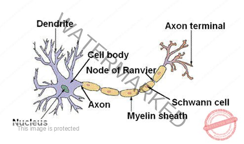
The outer cell membrane of a Schwann cell is called the neurilemma. The fact that axons in the
brain and spinal cord have no neurilemma is clinically significant because it plays an essential part in the regeneration of cut and injured axons.
Therefore the potential for regeneration in the brain and spinal cord is far less than it is in the peripheral nervous system.
Neuroglia
Neuroglia does not specialize in transmitting impulses.
Instead, they are special types of connective tissue cells.
Their name is appropriate because it is derived from Greek word glia meaning “glue.” One function of neuroglia cells is to hold the functioning neurons together and protect them.
Examples:
- Astrocytes: star-shaped cells, and are the main supporting tissue of the CNS.
- Epidymal cells: these cells form the epithelial lining of the ventricles and central canal of the spinal cord. Those cells form choroid plexuses that secrete cerebral spinal fluid.
- Microglia cells: these are derived from monocytes that migrate from blood into the nervous system before birth. They are found in the area of blood vessels. They enlarge and become phargocytic, removing microbes and damaged tissue in areas of inflammation and cell destruction.
- Oligodendrocytes: these are found in clusters around nerve cell bodies where they play a supportive function; form and maintain myelin, having the function as Schwann cells in peripheral nerves.
Impulse Generation and Conduction
The Nerve Impulse
The cell membrane of an unstimulated (resting) neuron carries an electric charge. Because of positive and negative ions concentrated on either side of the membrane, the inside of the membrane at rest is negative as compared with the outside.
A nerve impulse is a local reversal in the charge on the nerve cell membrane that then spreads along the membrane like an electric current. This sudden electrical change in the membrane is called an action potential. A stimulus, then, is any force that can start an action potential.
This electric change results from rapid shifts in sodium and potassium ions across the cell membrane: Na+ (usually extracellular, and K+ intracellular).
The reversal occurs very rapidly (in less than one thousandth of a second) and is followed by a rapid return of the membrane to its original state so that it can be stimulated again.
A myelinated nerve fiber conducts impulses more rapidly than an unmyelinated fiber of the same size because the electrical impulse “jumps” from node (space) to node in the myelin sheath instead of traveling continuously along the fiber.
The Synapse
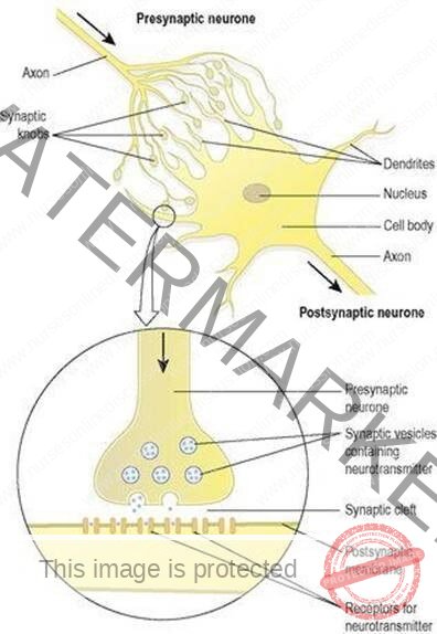
Each neuron is a separate unit, and there is no anatomic connection between neurons. How then is it possible for neurons to communicate? In other words, how does the axon of one neuron make functional contact with the membrane of another neuron? This is accomplished by the synapse, from a Greek word meaning “to clasp.” Synapses are points of junction for the transmission of nerve impulses.
Within the branching endings of the axon are small bubbles (vesicles) containing a type of chemical known as a neurotransmitter. When stimulated, the axon releases its neurotransmitter in to the narrow gap, the synaptic cleft, between the cells.
The neurotransmitter then acts as a chemical signal to stimulate the next cell, described as the postsynaptic cell. On the receiving membrane, usually that of a dendrite, sometimes another part of the cell, there are special sites, or receptors, ready to pick up and respond to specific neurotransmitters. Receptors in the cell membrane influence how or if that cell will respond to a given neurotransmitter.
Although there are many known neurotransmitters, the main ones are epinephrine, also called adrenaline; a related compound, norepinephrine, or noradrenaline; and acetylcholine. Acetylcholine (Ach) is the neurotransmitter released at the neuromuscular junction, the synapse between a neuron and a muscle cell.
All three of the above neurotransmitters function in the autonomic nervous system. It is common to think of neurotransmitters as stimulating the cells they reach; infact, they have been described as such in this discussion. Note, however, that some of these chemicals act to inhibit the postsynaptic cell and keep it from reacting.
Structure of a synapse: The axon ending has vesicles containing, neurotransmitter, which is released across the synaptic cleft to the membrane of the next cell
The Reflex Arc
As the nervous system functions, both external and internal stimuli are received, interpreted, and acted on. A complete pathway through the nervous system from stimulus to response is termed a reflex arc.
This is the basic functional pathway of the nervous system. The parts of a typical reflex arc are:
- Receptor-the end of a dendrite or some specialized receptor cell, as in a special sense organ, that detects a stimulus.
- Sensory neuron, or afferent neuron-a cell that transmits impulses toward the CNS.
- Central neuron-a cell or cells within the CNS. These neurons may carry impulses to and from the brain, may function within the brain, or may distribute impulses to different regions of the spinal cord.
- Motor neuron, or efferent neuron-a cell that carries impulses away from the CNS.
- Effectors- a muscle or a gland outside the CNS that carries out a response.
At its simplest, a reflex arc can involve just two neurons, one sensory and one motor, with a synapse in the CNS. There are very few reflex arcs that require only this minimal number of neurons. The knee jerk reflex is one of the few examples in humans. Most reflex arcs involve many more, even hundreds, of connecting neurons within the central nervous system.
Division of the Nervous System
The nervous system as a whole consists of two principal divisions called the central nervous system and peripheral nervous system. Because the brain and spinal cord occupy a midline or central location in the body, they are together called the central nervous system or CNS.
Similarly, the usual designation for the nerves of the body is the peripheral nervous system or PNS. Use of the term peripheral is appropriate because nerves extend to outlying or peripheral parts of the body.
A subdivision of the peripheral nervous system called the autonomic nervous system (ANS) structures that regulate the body’s autonomic or involuntary functions (for example, the heart rate, the contractions of the stomach, and intestines, and the secretion of chemical compounds by glands).
Central Nervous System
The CNS as its name implies, is centrally located. Its two major structures, the brain and spinal cord, are found along the midsagittal plane of the body. The brain is protected in the cranial cavity of the skull, and the spinal cord is surrounded in the spinal column. In addition, protective membranes called meninges cover the brain and spinal
Covering and Fluid Spaces Of the Brain and Spinal Cord
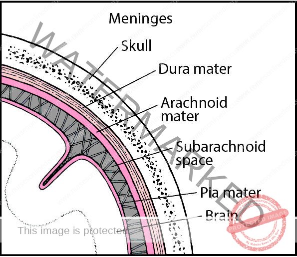
Nervous tissue is not a steady tissue. Even moderate pressure can kill nerve cells, so nature safeguards the chief organs made of this tissue-the spinal cord and the brain-by surrounding them with three fluid-containing membranes called the meninges.
The meninges are then surrounded by bone. The spinal meninges form tube like covering around the spinal cord and line the bony vertebral foramen of the vertebrae that surround the column. They are:
- the dura mater, which is the tough outer layer that lines the vertebral canal,
- the piamater, which is the innermost membrane covering the spinal cord itself,
- And the arachnoid, which is the membrane between the dura and pia mater.
The term arachnoid means “cobweblike.” It comes from arachne, which is the Greek word for spider. The meninges that form the protective covering around the spinal cord also extend up and around the brain to enclose it completely.
Flow of cerebral spinal fluid (diagram)
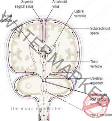
- Fluid fills the subarachnoid spaces between the pia mater and arachnoid in the brain and spinal cord. This fluid is called cerebrospinal fluid (CSF). Cerebrospinal fluid also fills spaces in the brain called cerebral ventricles.
- There are two lateral ventricles: One lies inside the right half of the cerebrum (the largest part of the human brain), and the other lies inside the left half of the cerebrum. The 3rd, and the 4th ventricles.
- CSF is one of the body’s circulating fluids. It forms continually from fluid filtering out of the blood in a network of brain capillaries known as the choroid plexus and into the ventricles.
- CSF seeps from the lateral ventricles into the third ventricle and flows down through the cerebral aqueduct into the fourth ventricle. It moves from the fourth ventricle into the small, tube like central canal of the cord and out into the subarachnoid spaces.
- Then it moves leisurely down and around the cord and up and around the brain (in the subarachnoid spaces of their meninges) and returns to the blood (in the veins of the brain)
Divisions of the Nervous system
CENTRAL NERVOUS SYSTEM
- Autonomic (involuntary) Nerves
- Brain, and
- Spinal cord
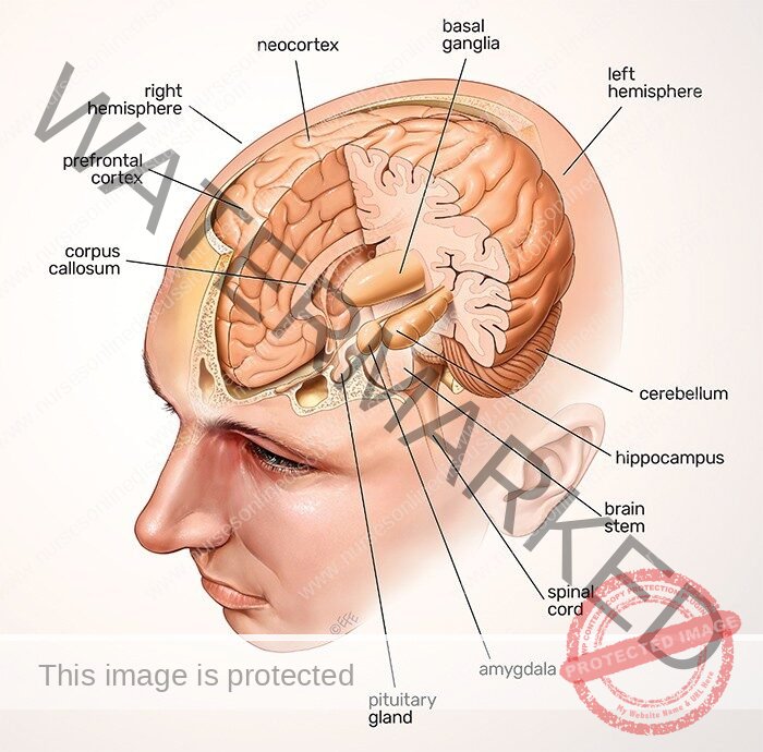
Divisions of the Brain
The brain, one of our largest organs, consists of the following major divisions, named in ascending order beginning with most inferior part:
Brain stem
- Medulla oblongata
- Pons
- Midbrain
Cerebellum
Ill. Diencephalon
- Hypothalamus
- Thalamus
- Cerebrum
Brain Stem
- The lowest part of the brain stem is the medulla oblongata.
- Immediately superior to the medulla lies the Pons and superior to that the midbrain. Together these three structures are called the brain stem.
- The medulla oblongata is an enlarged, upward extension of the spinal cord. It lies just inside the cranial cavity superior to the large hole in the occipital bone called the foramen magnum. Like the spinal cord, the medulla consists of gray and white matter, but their arrangement differs in the two organs.
- In the medulla, bits of gray matter mix closely and intricately with white matter to form the reticular formation (reticular means “netlike”). In the spinal cord, gray and white matter does not intermingle; gray matter forms the interior core of the cord, and white matter surrounds it. The Pons and midbrain, like the medulla, consist of white matter and scattered bits of gray matter.
- All three parts of the brain stem function as two-way conduction paths. Sensory fibers conduct impulses up from the cord to other parts of the brain, and motor fibers conduct impulses down from the brain to the cord. The cardiac, respiratory, and vasomotor centers (collectively called the vital centers) are located in the medulla.
- Impulses from these centers control heartbeat, respirations, and blood vessel diameter (which is important in regulating blood pressure). In addition, many important reflex centers lie in the brain stem.
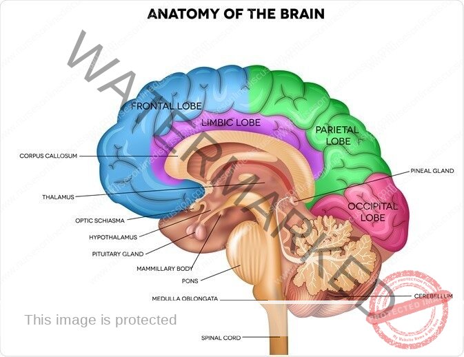
Diencephalon
The diencephalon is a small but important part of the brain located between the midbrain inferiorly and the cerebrum superiorly. It consists of two major structures: the hypothalamus and the thalamus. The ventricle of the diencephalons is the 3rd ventricle.
Hypothalamus:
- The hypothalamus, as its name suggests, is located inferior to the thalamus. The posterior pituitary gland, the stalk that attaches it to the undersurface of the brain, and areas of gray matter located in the sidewalls of a fluid-filled space called the third ventricle are extensions of the hypothalamus.
- Measured by size, it is one of the least significant parts of the brain, but measured its contribution to healthy survival; it is one of the most important brain structures. Impulses from neurons whose dendrites and cell bodies lie in the hypothalamus are conducted by their axons to neurons located in the spinal cord, and many of these impulses are then relayed to muscles and glands all over the body.
- Thus the hypothalamus exerts a major control over virtually all-internal organs. Among the vital functions that it helps control are the heartbeat, constriction and dilation of blood vessels, and contractions of the stomach and intestines.
- Some neurons in the hypothalamus function in a surprising way; they make the hormones that the posterior pituitary gland secretes into the blood.
- Because of one of these hormones (called antidiuretic hormone or ADH) affects the volume of urine excreted, the hypothalamus plays an essential role in maintaining the body’s water balance.
- Some of the neurons in the hypothalamus function as endocrine glands.
- Their axons secrete chemicals called releasing hormones into the blood, which then carries them to the anterior pituitary gland. Releasing hormones, as their name suggests, control the release of certain anterior pituitary hormones.
- These in turn influence the hormone secretion of other endocrine glands. Thus the hypothalamus indirectly helps control the functioning of every cell in the body.
- The hypothalamus is a crucial part of the mechanism for maintaining body temperature. Therefore a marked elevation in body temperature in the absence of disease frequently characterizes injuries or other abnormalities of the hypothalamus.
- In addition, this important center is involved in functions such as the regulation of water balance; sleep cycles, and the control of appetite and many emotions involved in pleasure, fear, anger, sexual arousal, and pain.
Thalamus
Just superior to the hypothalamus is a dumb-bell shaped section or largely gray matter called the thalamus.
Each enlarged end of the dumbbell lies in a lateral wall of the third ventricle. The thin center section of the thalamus passes from left to right through the third ventricle. The thalamus is composed chiefly of dendrites and cell bodies of neurons that have axons extending up to the sensory areas of the cerebrum. It performs the following functions:
- It helps produce sensations. Its neurons relay impulses to the cerebral cortex from the sense organ of the body.
- It associates sensations with emotions. Almost all sensations are accompanied by a feeling of some degree of pleasantness or unpleasantness. The way that these pleasant and unpleasant feelings are produced is unknown except that they seem to be associated with the arrival of sensory impulses in thalamus.
- It plays a part in the so -called arousal or alerting mechanism.
- It contains important nuclei such as medial geniculate which is responsible for auditory sense and lateral geniculate which is responsible for vision.
Cerebellum
The cerebellum is the second largest part of the human brain. It lies under the occipital lobe of the cerebrum.
In the cerebellum, gray matter composes the outer layer, and white matter composes the bulk of the interior.
Function. Most of our knowledge about cerebellar functions has come from observing patients who have some sort of disease of the cerebellum and from animals that have had the cerebellum removed.
From such observations, we know that the cerebellum plays an essential part in the production of normal movements.
Perhaps a few examples will make this clear. A patient who has a tumor of the cerebellum frequently loses balance and may topple over and feel like a drunken person when walking. It may be impossible to coordinate muscles normally. Frequent complaints about being clumsy and unable to even drive a nail or draw a straight line are typical.
With the loss of normal cerbellar functioning, the ability to make precise movements is lost. The general functions of the cerebellum, then, are to produce smooth coordinated movements, maintain equilibrium, and sustain normal postures.
Cerebrum
- The cerebrum is the largest and uppermost part of the brain. If you were to look at the outer surface of the cerebrum, the first features you would notice might be its many ridges and grooves. The ridges are called convolutions or gyri, and the grooves are called sulci.
- The deepest sulci are called fissures; the longitudinal fissure divides the cerebrum into right and left halves or hemispheres.
- These halves are almost separate structures except for their lower midportions, which are connected by a structure called the corpus callosum.
- Two deep sulci subdivide each cerebral hemisphere into four major lobes and each lobe into numerous convolutions.
- The lobes are named for the bones that lie over them: the frontal lobe, the parietal lobe, the temporal lobe, and the occipital lobe.
- A thin layer of gray matter, made up of neuron dendrites and cell bodies, composes the surface of the cerebrum. Its name is the cerebral cortex.
- White matter made up of bundles of neuronal fibers (tracts), composes most of the interior of the cerebrum. Within this white matter, however, are a few islands of gray matter known as the basal ganglia, whose functioning is essential for producing automatic movements and postures.
- Parkinson’s disease is a disease of the basal ganglia. Because shaking or tremors are common symptoms of Parkinson’s disease, it is also called “shaking palsy.”
- The nerve fibres of the white matter of the cerebral hemispheres are of three groups: commissural, association and projection.
Commissural fibres unite corresponding areas of the cortex of the two hemispheres across the midline. They comprise the corpus callosum, and the anterior and fornix commissures.
The corpus callosum is a broad band of fibres passing between corresponding cortical areas of the two hemispheres.
Association fibers – unite adjacent or widely separated gyri of the same hemisphere.
Projection fibers – ascend from or descend to lower lying parts of the central nervous system.
BRAIN AREA FUNCTION
Brain stem
Medulla oblongata Two-way conduction pathway between the spinal cord and higher brain centers; cardiac, respiratory, and vasomotor control center
Pons Two-way conduction pathway between areas of the brain and other regions of the body;
Influences respiration
Midbrain Two-way conduction pathway; relay for visual and auditory Impulses
Diencephalon
Hypothalamus Regulation of body temperature, water balance, sleeps cycle control appetite, and sexual arousal
Thalamus Sensory relay station from various body areas to cerebral cortex; emotions and alerting or arousal mechanisms
Cerebellum Muscle coordination; maintenance of equilibrium and posture
Cerebrum Sensory perception, emotions willed movements, consciousness, and memory
Functions of the cerebral cortex
It’s within the cerebral cortex that nerve impulses are received and analyzed. These activities form the basis of knowledge. The brain “stores” information, much of which can be recalled on demand by means of a phenomenon called memory. It’s in the cortex that thought processes such as association, judgment, and discrimination takes place. Conscious deliberations and voluntary actions also arise from there.
Although the various brain areas act in coordination to produce behavior, particular functions are localized in the cortex of each lobe:
Draw the diagram of functional areas
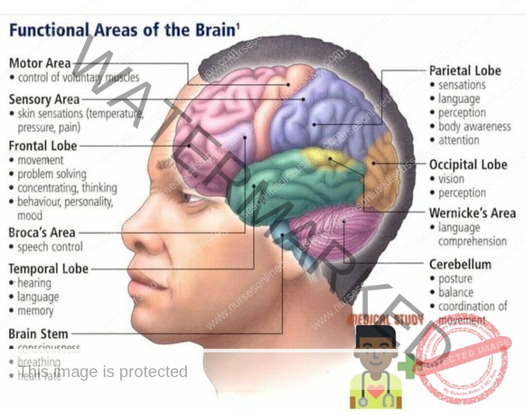
- The frontal lobe: this is relatively larger in humans than any other organism, lies anterior to the central sulcus. This lobe contains a primary motor area, which provides conscious control of the skeletal muscles. A larger region of the lobe is involved in planning and conscious thought. It also contains two areas important in speech.
- The parietal lobe: occupies the superior part of each hemisphere and lies posterior to the central sulcus. A large region of the parietal lobe integrates information from all sensory areas. The estimation of distances, sizes, and shapes also take place here.
The gyrus just behind the central sulcus contains the primary sensory area, where impulses from the skin, such as touch, pain, and temperature are interpreted. As with the motor cortex, the greater the intensity of sensation from a particular area, the tongue or fingers, for example, the more area of the cortex is involved.
- The temporal lobe: lies inferior to the lateral sulcus and folds under the hemisphere on each side. This lobe contains the auditory areas for receiving and interpreting impulses from the ear. The olfactory area, concerned with the sense of smell, is located in the medial part of the temporal lobe and is not visible from the surface; it’s stimulated by impulses arising from receptors in the nose.
- The occipital lobe: lies posterior to the parietal lobe and extend over the cerebellum. This lobe contains for interpreting impulses arising from the retina of the eye.
Communication areas
- The ability to communicate by written and verbal means is an interesting example of how areas of the cerebral cortex are interrelated. The intergration of these areas is closely connected with the learning process.
- The auditory receiving area: detects sound impulses transmitted from the environment, where as the surrounding area; the association area, interprets the sounds such as; integrating the pitch, loudness, and direction of sound. Another area is located in the left side in most people; the comprehension area, or wernick area. This area functions in speech recognition and the meaning of words. Someone who suffers damage in this area of the brain, as by stroke, will have difficulty in understanding the meaning of speech.
- The beginnings of language are learned by hearing; thus, the auditory area for understanding sounds is near the auditory receiving areas of the cortex. Babies often appear to understand what is being said long before they do any talking themselves. It is several years before the children learn to read or write words.
- The visual areas of the occipital cortex: visual receiving area; visual images of language are collected. The visual association area that lies anterior interprets these visual impulses as words. The ability to read with understanding also develops in this area.
- The motor areas for spoken and written communication: lie anterior to the most inferior part of the frontal lobe’s motor cortex. The specialized cortical region; motor speech area, or Broca area, plans the sequence of muscle contractions in the tongue, larynx, and soft palate required to form meaningful sentences.
- There is a functional relation among areas of the brain. Many neurons must work together to enable to enable a person to receive, interpret, and respond to verbal and written messages as well as to touch and other sensory stimuli.
Memory and the learning process
- Memory is the mental faculty for recalling ideas. Short-term memory refers to the retention of bits of information for a few seconds or perhaps a few minutes, after which the information for a few minutes after which it’s lost unless reinforced.
- Long-term memory refers o the storage of information that can be recalled at a later time. There is a tendency for a memory to become more fixed the more often the person repeats the remembered experience; thus short term memory signals can lead to long term memories.
- Physiological studies show that rehearsal of the same information again and again accelerates the degree of short term memory transfer into long term memory. It’s also noted that the brain is able to organize information so that new ideas are stored in the same areas in which similar ones have been stored before.
- Along the border between the cerebrum and the diencephalon is a region known as the limbic system. This system is involved in emotional states and behavior. It’s also important for short term memories and the consolidation of long term memories, and thus is essential for learning.
Spinal Cord
Location of the Spinal Cord
In the embryo, the spinal cord occupies the entire spinal canal and so extends down into the tail portion of the vertebral column. However, the column of bone grows much more rapidly than the nerve tissue of the cord, so that eventually the end of the cord no longer reaches the lower part of the spinal canal. This disparity in growth continues to increase; in the adult the cord ends in the region just below the area to which the last rib attaches (between the first and the second lumbar vertebrae.
Structure of the Spinal Cord
- The spinal cord lies within the vertebral canal and extends from the foramen magnum to the level of the second lumbar vertebrae after which a fibrous remnant, the filum terminale, descends to be attached to the back of the coccyx. The cord is about 45 cm long. It is cylindrical in shape, flattened slightly anteroposteriorly, and has cervical and lumbar enlargements where the nerves supplying the upper and lower limb originate.
- Since the spinal cord is shorter than the vertebral canal, the nerves descend with increasing obliquity before leaving the canal through the intervertebral foramina.
- The collection of lower lumbar, sacral and coccygeal nerves below the spinal cord, with the filum terminale, is known as the cauda equina.
- The cord has an anterior median fissure and a posterior median sulcus. On its sides the rootlets of the spinal nerves emerge from anterolateral and posterolateral sulci.
- The spinal cord has a small, irregular shaped internal section that consists of gray matter (nerve cell bodies) and a larger area surrounding this gray part that consists of white matter (nerve cell fibers).
- The gray matter is so arranged that a column of cells extend up and down dorsally, one on each side; another column is found in the central region on each side. These two pairs of columns, called the dorsal and ventral horns, give the gray matter an H-shaped appearance in cross section.
- In the center of the gray matter is a small channel, central canal that contains cerebrospinal fluid, the liquid that circulates around the brain and spinal cord. The white matter consists of thousands of nerve cell fibers arranged in three areas external to the gray matter on each side.
Functions of the Spinal Cord
The spinal cord is the link between the spinal nerves and the brain. It is also a place where simple responses, known as reflexes can be coordinated even without involving the brain.
The functions of the spinal cord may be divided into three categories;
- Conduction of sensory impulses upward through ascending tracts to the brain
- Conduction of motor impulses from the brain down through descending tracts to the efferent neurons that supply muscles or glands
- Reflex activities. A reflex is a simple, rapid, and automatic response involving very few neurons.
When you fling out an arm or leg to catch your balance, withdraw from a painful stimulus, or blink to avoid an object approaching your eyes, you are experiencing reflex behavior. A reflex pathway that passes through the spinal cord alone and does not involve the brain is termed a spinal reflex. The stretch reflex, in which a muscle is stretched and responds by contracting, is one example.
If you tap the tendon below the kneecap (the patellar tendon): the muscles of the anterior thigh (quadriceps femoris) contracts, eliciting the knee jerk. Such stretch reflexes may be evoked by appropriate tapping of most large muscles (such as the triceps brachii in the arm and the gastrocnemius in the calf of the leg).
Because reflexes occur automatically, they are used in physical examinations to test the condition of the nervous system.
Peripheral Nervous System
The nerves connecting the brain and the spinal cord to other parts of the body constitute the peripheral nervous system (PNS). This system includes cranial and spinal nerves that connect the brain and spinal cord, respectively, to peripheral structures such as the skin surface and the skeletal muscles.
In addition, other structures in the autonomic nervous system (ANS) are considered part of the PNS. These connect the brain and spinal cord to various glands in the body and to the cardiac and smooth muscle in the thorax and abdomen.
Base of the brain showing cranial nerve (revise this from your text book)
Nerves
- A nerve is a bundle of nerve cell fibers located outside the CNS. Bundles of nerve cell fibers within the CNS are tracts.
- Tracts are located within the brain and also within the spinal cord to conduct impulses to and from the brain. A nerve or tract can be compared to an electric cable made up of many wires. As with muscles, the “wires,” or nerve cell fibers in a nerve, are bound together with connective tissue.
- A few of the cranial nerves have only sensory fibers for conducting impulses toward the brain. These are described as sensory, or afferent, nerves. A few of the cranial nerves contain only motor fibers for conducing impulses away from the brain and are classified as motor, or efferent, nerves.
- However, the remainder of the cranial nerves and all of the spinal nerves contain both sensory and motor fibers and are referred to as mixed nerves.
Location of the Cranial Nerves
Cranial nerves are nerves that are attached to the brain.
There are 12 pairs of cranial nerves (henceforth, when a cranial nerve is identified, a pair is meant). They are numbered according to their connection with the brain; beginning at the front and proceeding back. The first 9 pairs and the 12th pair supply structures in the head.
General Functions of the cranial nerves
From the functional point of view, we may think of the kinds of messages the cranial nerves handle as belonging to one of four categories:
- Special sensory impulses, such as for smell, taste, vision, and hearing
- General sensory impulses, such as those for pain, touch, temperature, deep muscle sense, pressure, and vibrations
- Somatic motor impulses resulting in voluntary control of skeletal muscles
- Viscera motor impulses producing involuntary control of glands and involuntary muscles (cardiac and smooth muscle). These motor pathways are part of the autonomic nervous system, parasympathetic division.
Names and Functions of the Cranial Nerves
The 12 cranial nerves are always numbered according to the traditional Roman style:
- cranial nerves−I, II, and VIll−contain only sensory fibers;
- Cranial nerve −III, IV, VI, XI and Xll− contain all or mostly motor fibers.
The remainder−V, VII, IX, and X− contain both sensory and motor fibers; they are known as mixed nerves.
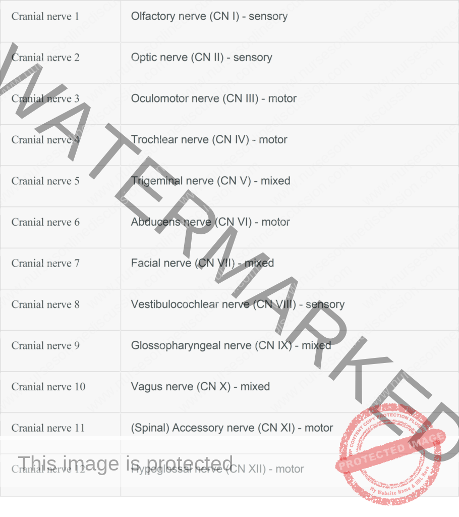
All 12 nerves are listed below
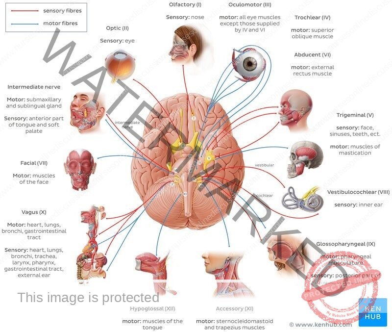
- The olfactory nerve they supply the olfactory mucous membrane in the upper part of the nasal cavity. The nerve fibers originate I the bipolar olfactory cells of the mucosa and join to form 15-20 olfactory bundles which pass through the cribriform plate of the ethmoid bone to reach the olfactory bulb.
- The optic nerve it is the sensory nerve of the retina. Its fibres originate in the ganglion layer and converge on the posterior part of the eye ball. The nerve passes backwards through the orbit and optic canal into the middle cranial fossa where it unites with the nerve of opposite side of the optic chiasma.
- The oculomotor nerve this nerve has somatic motor and general visceral (parasympathetic) motor fibres.
The somatic fibres supply the bulbar muscles, except superior oblique and lateral rectus. The parasympathetic fibres synapse in the ciliary ganglion and supply the sphincter pupillae and ciliary muscle.
The nuclei of the nerve are situated in the upper midbrain in the peri-aqueductal grey matter. The nerve fibres pass forwards through the midbrain and leave it between the cerebral peduncles. The nerve pass through the posterior and middle cranial fossae and divides into superior and inferior divisions near the superior orbital fissure.
- The trochlear nerve this is the somatic motor nerve supply to the superior oblique. Its nucleus lies in the lower midbrain in the peri-aqueductal grey matter.
The fibres pass posteriorly and undergo a dorsal decussation with the nerve of the opposite side caudal to the inferior colliculi the nerve then passes forwards through the posterior and middle cranial fossae, enters the orbit through the superior orbital fissure and supplies superior oblique.
- The trigeminal nerve is the great sensory nerve of the face and head. It has three branches that transport general sense impulses (e.g., pain, touch, temperature) from the eye, the upper jaw, and the lower jaw. The third branch is joined by motor fibers to the muscles of mastication (chewing).
- The abducens nerve is a somatic motor nerve supplying lateral rectus. Its nucleus is situated in the lower pons. The nerve leaves the inferior border of the pons near the midline, passes forwards through the posterior and middle cranial fossae, the cavernous sinus and the orbit, and supplies lateral rectus.
- The facial nerve is largely motor; Branches from the facial nerve supply all the muscles of facial expression.
This nerve also includes special sensory fibers for taste (anterior two thirds of the tongue), and it contains secretary fibers to the smaller salivary glands (the submandibular and sublingual) and to the lacrimal gland.
- The vestibulocochlear nerve contains special sensory fibers for hearing as well as those for balance from the semi circular canals of the internal ear. This nerve is also called the auditory or acoustic nerve.
- The glossopharyngeal nerve contains general sensory fibers from the back of the tongue and the pharynx (throat). This nerve also contains sensory fibers for taste from the posterior third of the tongue, secretary fibers that supply the largest salivary gland (parotid), and motor nerve fibers to control the swallowing muscles in the pharynx.
- The vagus nerve is the longest cranial nerve. (Its name means “wanderer. “) It supplies most of the organs in the thoracic and abdominal cavities. This nerve also contains motor fibers to the larynx (voice box) and pharynx, and to glands that produce digestive juices and other secretions.
- The accessory nerve (formerly called the spinal accessory nerve) is a motor nerve with two branches.
One branch controls two muscles of the neck, the trapezius and sternocleidomastoid ; the other supplies muscles of the larynx
- The hypoglossal nerve, the last of the 12 cranial nerves, carries impulses controlling the muscles of the tongue.
Spinal Nerves
Location and Structure of Spinal Nerves
- Spinal nerves arise from spinal cord. There are 31 pairs of spinal nerves, each pair numbered according to the level of the spinal cord from which it arises. Each nerve is attached to the spinal cord by two roots; the dorsal root and the ventral root.
- The roots are formed from a number of rootlets which emerge from the anterolateral and posterolateral sulci of the spinal cord. The ventral root carries efferent (motor) fibres from the cord and the dorsal root, afferent (sensory) fibres to the cord. The cell bodies of the sensory fibres are situated in a ganglion on the dorsal root.
- The spinal nerves are therefore a mixture of motor and sensory fibres. Each nerve leaves the vertebral canal through an intervertebral foramen and soon divides into a large ventral and smaller dorsal ramus (branch).
- The adjacent ventral rami of most regions communicate to form plexuses (cervical, brachial and lumbo-sacral) while those of the thoracic region become the intercostals and subcostal nerves. The dorsal rami pass backwards into the postvertebral muscles and divide into medial and lateral branches.
- These rami supply the muscles and skin over the posterior aspect of the body but give no branches to the limbs. The ventral rami supply the anterior and lateral wall of the back and the lower limbs.
Branches of the Spinal Nerves
Each spinal nerve continues only a very short distance away from the spinal cord and then branches into small posterior divisions and rather large anterior divisions. The larger anterior branches interlace to form networks called plexuses, which then distribute branches to the body parts. The three main plexuses are described as follows:
- The cervical plexuses supplies motor impulses to the muscles of the neck and receive sensory impulses from the neck and the back of the head. The phrenic nerve, which activates the diaphragm, arises from this plexus.
- The brachial plexus sends numerous branches to the shoulder, arm, forearm, wrist, and hand. The radial nerve emerges from the brachial plexus.
- The lumbosacral plexus supplies nerves to the lower extremities. The largest of these branches is the sciatic nerve, which leaves the dorsal part of the pelvis, passes beneath the gluteus maximus muscle, and extends down the back of the thigh. At its beginning it is nearly 1 inch thick, but it soon branches to the thigh muscles; near the knee it forms two subdivisions that supply the leg and the foot.
The Autonomic Nervous System
Parts of the Autonomic Nervous System
Although the internal organs such as the heart, lungs, and stomach contain nerve endings and nerve fibers for conducting sensory messages to the brain and cord, most of these impulses do not reach consciousness.
These afferent impulses from the viscera are translated into reflex responses without reaching the higher center of the brain; the sensory neurons from the organs are grouped with those that come from the skin and voluntary muscles.
In contrast, the efferent neurons, which supply the glands and the involuntary muscles, are arranged very differently from those that supply the voluntary muscles.
This variation in the location and arrangement of the visceral efferent neurons has led to their classification as part of a separate division called the autonomic nervous system (ANS). The ANS itself is comprised of sympathetic and parasympathetic divisions.
The autonomic nervous system has many ganglia that serve as relay stations. In these ganglia each message is transferred at a synapse from the first neuron to a second one and from there to the muscle or gland cell.
This differs from the voluntary (somatic nervous system, in which each motor nerve fiber extends all the way from the spinal cord to the skeletal muscle with no intervening synapse.
Some of the autonomic fibers are within the spinal nerves; some are within the cranial nerves. The distribution of the two divisions of the ANS is as follows:
- The sympathetic pathways begin in the spinal cord with cell bodies in the thoracic and lumbar regions, the thoracolumbar area. The sympathetic fibers arise from the spinal cord at the level of the first thoracic nerve down to the level of the second lumbar spinal nerve. From this part of the cord, nerve fibers extend to ganglia where they synapse with a second set of neurons, the fibers of which extend to the glands and involuntary muscle tissues.
Many of the sympathetic ganglia form the sympathetic chains, two cords- like strands of ganglia that extend along either side of the spinal column from the lower neck to the upper abdominal region. The nerves that supply the organs of the abdominal and pelvic cavities synapse in three single ganglia farther from the spinal cord.
The second neurons of the sympathetic nervous system acton the effectors by releasing the neurotransmitter epinephrine (adrenaline). This system is therefore \ described as adrenergic, which means “activated by adrenaline.”
- The parasympathetic pathways begin in the craniosacral areas, with fibers arising from cell bodies of the midbrain, medulla, and lower (sacral) part of the spinal cord. From these centers the first set of fibers extends to autonomic ganglia that are usually located near or within the walls of the effector organs.
The pathways then continue along a second set of neurons that stimulate the involuntary tissues. These neurons release the neuro transmitter acetylcholine, leading to the description of this system as cholinergic (activated by acetylcholine).
Functions of the Autonomic Nervous System
The autonomic nervous system regulates the action of the glands, the smooth muscles of the hollow organs and vessels, and the heart muscle. These actions are all carried on automatically; whenever any changes occur that call for a regulatory adjustment, the adjustment is made without conscious awareness.
The sympathetic part of the autonomic nervous system tends to act as an accelerator for those organs needed to meet a stressful situation. It promotes what is called the fight-or-flight response. If you think of what happens to a person who is frightened or angry, you can easily remember the effects of impulses from the sympathetic nervous system:
- Stimulation of the central portion of the adrenal gland. This produces hormones, including epinephrine, that prepare the body to meet emergency situations in many ways. The sympathetic nerves and hormones from the adrenal gland reinforce each other.
- Dilation of the pupil and decrease in focusing ability (for near objects)
- Increase in the rate and force of heart contractions
- Increase in blood pressure due partly to the more effective heartbeat and partly to constriction of small arteries in the skin and the internal organs
- Dilation of blood vessels to skeletal muscles, bringing more blood to these tissues
- Dilation of the bronchial tubes to allow more oxygen to enter
- Increase in metabolism.
The sympathetic system also acts as a brake on those systems not directly involved in the response to stress, such as the urinary and digestive systems. If you try to eat while you are angry, you may note that your saliva is thick and so small in amount that you can swallow only with difficulty.
Under these circumstances, when food does reach the stomach, it seems to stay there longer than usual.
The parasympathetic part of the autonomic nervous system nonnal1y acts as a balance for the sympathetic system once a crisis has passed. The parasympathetic system brings about constriction of the pupils, slowing of the heart rate, and constriction of the bronchial tubes.
It also stimulates the formation and release of urine and activity of the digestive tract.
Saliva, for example, flows more easily and profusely and its quantity and fluidity increase.
Most organs of the body receive both sympathetic and parasympathetic stimulation, the effects of the two systems on a given organ generally being opposite.
We are a supportive platform dedicated to empowering student nurses and midwives through quality educational resources, career guidance, and a vibrant community. Join us to connect, learn, and grow in your healthcare journey
Quick Links
Our Courses
Legal / Policies
Get in Touch
(+256) 790 036 252
(+256) 748 324 644
Info@nursesonlinediscussion.com
Kampala ,Uganda
© 2026 Nurses online discussion. All Rights Reserved