Urinary System
Subtopic:
Renal Failure

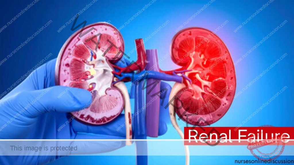
Renal failure, also known as kidney failure, is a medical condition in which the kidneys are unable to adequately filter waste products and excess fluid from the blood. This leads to a buildup of toxins and fluid in the body, which can cause a range of health problems.
The kidneys play vital roles in maintaining homeostasis, including filtering blood, producing urine, regulating blood pressure, producing erythropoietin (a hormone that stimulates red blood cell production), and maintaining electrolyte and acid-base balance. When these functions are impaired, the entire body is affected.
Functions of the Kidneys
Before delving into renal failure, it’s essential to understand the critical functions of healthy kidneys:
Filtration: The kidneys filter blood to remove waste products, toxins, and excess water, forming urine.
Reabsorption: Essential substances like glucose, amino acids, water, and electrolytes are reabsorbed back into the bloodstream.
Secretion: Waste products and excess ions are secreted from the blood into the renal tubules for excretion in urine.
Hormone Production:
Erythropoietin: Stimulates the bone marrow to produce red blood cells.
Renin: Involved in regulating blood pressure (Renin-Angiotensin-Aldosterone System – RAAS).
Calcitriol (Active Vitamin D): Essential for calcium and phosphate absorption and bone health.
Blood Pressure Regulation: Kidneys regulate blood pressure through the RAAS and by controlling fluid volume.
Electrolyte Balance: Kidneys maintain the balance of electrolytes such as sodium, potassium, calcium, and phosphate.
Acid-Base Balance: Kidneys help regulate the body’s pH by excreting excess acids or bases.
When kidneys fail, these functions are compromised, leading to systemic effects.
Types of Renal Failure
Renal failure is broadly classified into two main types:
Acute Kidney Injury (AKI): A sudden and often reversible loss of kidney function.
Chronic Kidney Disease (CKD): A progressive and irreversible decline in kidney function over months or years.
Acute Kidney Injury (AKI)
AKI is characterized by a rapid decrease in kidney function, typically occurring over hours or days. It leads to a buildup of nitrogenous waste products (like urea and creatinine) and fluid and electrolyte imbalances. AKI is often a complication of other severe illnesses.
Causes/Etiology of AKI:
AKI causes are categorized based on where the problem originates:
Prerenal AKI: Caused by decreased blood flow to the kidneys (perfusion). The kidney structure is initially intact.
Examples: Severe dehydration, hemorrhage, heart failure, sepsis, severe burns, excessive diuretic use, renal artery stenosis.
Intrarenal AKI: Caused by direct damage to the kidney tissue itself.
Examples:
Acute Tubular Necrosis (ATN): The most common cause, often due to prolonged prerenal ischemia or nephrotoxic substances (certain antibiotics, contrast dyes, NSAIDs).
Acute Interstitial Nephritis: Allergic reaction to medications (e.g., penicillin, NSAIDs, sulfa drugs) or infections.
Glomerulonephritis: Inflammation of the glomeruli.
Vascular Disorders: Vasculitis, thrombotic microangiopathies.
Postrenal AKI: Caused by an obstruction of urine flow from the kidneys.
Examples: Kidney stones (bilateral or in a single functioning kidney), enlarged prostate (BPH), tumors (bladder, prostate, pelvic), strictures in the ureters or urethra, neurogenic bladder.
Pathophysiology of AKI:
The pathophysiology varies depending on the cause:
Prerenal: Reduced renal blood flow leads to decreased glomerular filtration rate (GFR). The kidneys attempt to compensate by reabsorbing more sodium and water, leading to concentrated urine. If ischemia is prolonged, it can lead to tubular cell damage (ATN).
Intrarenal: Damage to the nephrons (glomeruli, tubules, interstitium, or blood vessels) impairs filtration, reabsorption, and secretion. ATN involves damage and death of tubular cells, leading to sloughing and obstruction of tubules, further reducing GFR.
Postrenal: Obstruction of urine outflow causes a backup of pressure in the collecting system and renal tubules, which reduces GFR. Prolonged obstruction can lead to kidney damage.
Clinical Manifestations of AKI:
Symptoms often depend on the underlying cause and the severity of kidney dysfunction.
Oliguria (urine output < 400 ml/day) or Anuria (urine output < 100 ml/day): Common, especially in intrinsic and postrenal AKI. Prerenal AKI may initially have low but concentrated urine output.
Fluid Overload: Peripheral edema, pulmonary edema (shortness of breath, crackles), weight gain, elevated blood pressure.
Electrolyte Imbalances:
Hyperkalemia (muscle weakness, ECG changes, arrhythmias)
Hyponatremia (confusion, seizures)
Hyperphosphatemia
Hypocalcemia
Acid-Base Imbalance: Metabolic acidosis (Kussmaul respirations).
Uremia (buildup of waste products): Fatigue, weakness, nausea, vomiting, loss of appetite, confusion, pruritus (itching), metallic taste in mouth.
Other: Flank pain (if obstruction), fever (if infection), rash (if allergic reaction).
Diagnosis of AKI:
History and Physical Examination: Identifying potential causes (dehydration, medications, obstruction symptoms).
Laboratory Tests:
Serum Creatinine and Blood Urea Nitrogen (BUN): Elevated levels indicate decreased kidney function. A rapid rise is characteristic of AKI.
Serum Electrolytes: To detect imbalances (potassium, sodium, phosphate, calcium).
Arterial Blood Gases (ABGs): To assess acid-base status.
Complete Blood Count (CBC): May show anemia (if prolonged AKI) or signs of infection.
Urine Tests:
Urinalysis: May show protein, blood, casts (cellular debris), or signs of infection.
Urine Output Measurement: Monitoring hourly and daily output is crucial.
Fractional Excretion of Sodium (FENa): Helps differentiate prerenal from intrarenal AKI.
Imaging Studies:
Renal Ultrasound: To assess kidney size, look for obstruction (hydronephrosis), and rule out chronic kidney disease.
CT scan or MRI: May be used to identify causes of obstruction or other kidney abnormalities.
Kidney Biopsy: May be necessary to determine the specific cause of intrarenal AKI (e.g., glomerulonephritis, interstitial nephritis).
Management of AKI:
Management focuses on treating the underlying cause, preventing further kidney damage, managing complications, and supporting kidney function.
Treat the Underlying Cause:
Restore blood flow in prerenal AKI (fluid resuscitation, improving cardiac function).
Remove offending agents (nephrotoxic drugs).
Relieve obstruction in postrenal AKI (catheterization, stenting, surgery).
Treat infections or inflammatory conditions.
Fluid and Electrolyte Management:
Careful fluid balance monitoring. Fluid restriction if fluid overloaded.
Management of hyperkalemia (medications like IV calcium gluconate, insulin and glucose, sodium polystyrene sulfonate, or dialysis).
Correction of hyponatremia, hyperphosphatemia, hypocalcemia.
Medication Management:
Adjusting dosages of renally excreted medications.
Avoiding nephrotoxic drugs.
Diuretics may be used to manage fluid overload, but their effectiveness in AKI is limited.
Nutritional Support:
Protein restriction may be necessary to reduce nitrogenous waste production.
Adequate calorie intake.
Restriction of potassium and phosphate intake.
Renal Replacement Therapy (Dialysis): May be needed if AKI is severe and complications like severe fluid overload, refractory hyperkalemia, severe metabolic acidosis, or uremic symptoms are present.
Hemodialysis
Peritoneal Dialysis
Continuous Renal Replacement Therapy (CRRT)
Nursing Management of AKI:
Monitor Vital Signs: Blood pressure, heart rate, respiratory rate, temperature.
Monitor Fluid Balance: Accurate intake and output (I&O) measurement, daily weight. Assess for signs of fluid overload or dehydration.
Monitor Laboratory Results: BUN, creatinine, electrolytes, ABGs.
Assess for Signs of Complications: Changes in mental status (uremia), signs of infection, bleeding, cardiac arrhythmias (hyperkalemia), respiratory distress (pulmonary edema).
Medication Administration: Administer medications as prescribed, adjusting dosages as needed. Avoid nephrotoxic drugs.
Nutritional Support: Provide prescribed diet, monitor intake, assess for nausea/vomiting.
Skin Care: Assess for pruritus and provide skin care to prevent breakdown.
Patient and Family Education: Explain the condition, treatment plan, fluid/dietary restrictions, and signs/symptoms to report.
Emotional Support: Provide support to the patient and family dealing with a critical illness.
Chronic Kidney Disease (CKD)
CKD is a long-term condition characterized by a gradual and irreversible loss of kidney function over months to years. It is defined by kidney damage or decreased kidney function for three months or more, regardless of the cause. CKD is staged based on the estimated glomerular filtration rate (eGFR).
Causes/Etiology of CKD:
The most common causes of CKD are:
Diabetes Mellitus: Diabetic nephropathy is the leading cause. High blood glucose damages the small blood vessels in the kidneys.
Hypertension: High blood pressure damages the kidney’s blood vessels and filtering units.
Glomerulonephritis: Inflammation of the glomeruli.
Polycystic Kidney Disease (PKD): A genetic disorder causing cysts to grow in the kidneys.
Interstitial Nephritis: Inflammation of the kidney tubules and surrounding tissue.
Obstructive Uropathy: Long-standing obstruction of urine flow (e.g., enlarged prostate, kidney stones, strictures).
Recurrent Kidney Infections (Pyelonephritis).
Certain Medications: Long-term use of certain drugs like NSAIDs.
Pathophysiology of CKD:
Regardless of the initial cause, the progressive damage in CKD involves the loss of functioning nephrons. As nephrons are destroyed, the remaining healthy nephrons hypertrophy (enlarge) and work harder to compensate (hyperfiltration). This compensatory mechanism can maintain kidney function for a time, but eventually, the increased workload damages the remaining nephrons, leading to a vicious cycle of progressive decline. This leads to a decrease in GFR and the kidneys’ inability to perform their functions, resulting in:
Accumulation of waste products (uremia).
Fluid and electrolyte imbalances (sodium and water retention, hyperkalemia, hyperphosphatemia, hypocalcemia).
Metabolic acidosis.
Anemia (decreased erythropoietin production).
Bone disease (renal osteodystrophy) due to impaired calcium and phosphate metabolism and decreased active Vitamin D production.
Hypertension (due to fluid overload and RAAS activation).
Cardiovascular disease (a major complication and cause of death in CKD).
Clinical Manifestations of CKD:
Symptoms often do not appear until kidney function is significantly impaired (usually < 20-25% of normal function). Symptoms are often non-specific and affect multiple body systems due to uremia.
Urinary Changes: May vary depending on the stage. Early stages may have no changes or polyuria (especially in diabetic nephropathy). As CKD progresses, oliguria or anuria may occur. Proteinuria (protein in urine) and hematuria (blood in urine) may be present.
Fluid Overload: Peripheral edema, pulmonary edema, weight gain, shortness of breath, hypertension.
Electrolyte Imbalances:
Hyperkalemia (muscle weakness, arrhythmias).
Hyponatremia (often dilutional due to fluid overload).
Hyperphosphatemia (itching, bone pain).
Hypocalcemia (muscle cramps, tingling).
Cardiovascular System: Hypertension, heart failure, pericarditis, arrhythmias, atherosclerosis.
Hematologic System: Anemia (fatigue, weakness, pallor), bleeding tendencies (platelet dysfunction).
Gastrointestinal System: Nausea, vomiting, anorexia, metallic taste, stomatitis, constipation or diarrhea.
Neurological System: Fatigue, weakness, difficulty concentrating, confusion, peripheral neuropathy (restless legs syndrome, paresthesias), muscle cramps, seizures, uremic encephalopathy in advanced stages.
Musculoskeletal System: Renal osteodystrophy (bone pain, fractures, muscle weakness), joint pain.
Integumentary System: Pruritus (itching), dry skin, uremic frost (rare, crystal deposits on skin), pallor, yellowish-brown discoloration.
Endocrine/Metabolic: Impaired glucose metabolism, hyperlipidemia, gout.
Diagnosis of CKD:
History and Physical Examination: Identifying risk factors (diabetes, hypertension) and symptoms.
Laboratory Tests:
Serum Creatinine and BUN: Persistently elevated levels.
Estimated Glomerular Filtration Rate (eGFR): Calculated from serum creatinine, age, sex, and race. Used to stage CKD.
Serum Electrolytes: To detect imbalances.
Albumin-to-Creatinine Ratio (ACR) or Proteinuria: Elevated levels indicate kidney damage.
CBC: To assess for anemia.
Serum Calcium, Phosphate, Parathyroid Hormone (PTH), Vitamin D: To assess for renal osteodystrophy.
Lipid Profile: To assess for hyperlipidemia.
Urine Tests: Urinalysis, 24-hour urine collection for protein and creatinine clearance.
Imaging Studies: Renal Ultrasound to assess kidney size (often small and scarred in advanced CKD, but may be normal or enlarged in PKD or obstructive causes), look for obstruction.
Kidney Biopsy: May be done to determine the specific cause of CKD, especially if the cause is unclear or if considering specific treatments.
Management of CKD:
The goals of CKD management are to slow the progression of kidney damage, manage complications, and prepare for renal replacement therapy if needed.
Manage Underlying Causes: Strict control of blood glucose in diabetes, strict blood pressure control (often using ACE inhibitors or ARBs which also protect the kidneys), managing glomerulonephritis or other specific causes.
Dietary Management:
Protein Restriction: To reduce the buildup of nitrogenous waste products.
Sodium Restriction: To help control blood pressure and fluid retention.
Potassium Restriction: To prevent hyperkalemia.
Phosphate Restriction: To manage hyperphosphatemia. Phosphate binders are often prescribed with meals.
Fluid Restriction: May be necessary in later stages if fluid overloaded.
Medication Management:
Antihypertensives: ACE inhibitors, ARBs, calcium channel blockers, diuretics.
Phosphate Binders: Taken with meals to reduce phosphate absorption (e.g., calcium carbonate, sevelamer).
Vitamin D Supplements: Active Vitamin D (calcitriol) to help with calcium absorption and bone health.
Erythropoiesis-Stimulating Agents (ESAs): To treat anemia (e.g., epoetin alfa, darbepoetin alfa). Iron supplements are often needed as well.
Diuretics: To manage fluid overload.
Medications to manage hyperkalemia: If dietary restriction is insufficient.
Management of Complications:
Treat cardiovascular risk factors.
Manage bone disease.
Treat metabolic acidosis (sodium bicarbonate).
Preparation for Renal Replacement Therapy (RRT): As CKD progresses to End-Stage Renal Disease (ESRD), RRT becomes necessary.
Dialysis: Hemodialysis or Peritoneal Dialysis.
Kidney Transplantation: A preferred option for eligible patients.
Palliative Care: For patients who choose not to pursue RRT or for whom it is not suitable, focus shifts to symptom management and comfort.
Nursing Management of CKD:
Monitor Vital Signs: Blood pressure is crucial.
Monitor Fluid Balance: Accurate I&O, daily weight. Assess for signs of fluid overload.
Monitor Laboratory Results: Trends in BUN, creatinine, eGFR, electrolytes, CBC, calcium, phosphate, PTH.
Assess for Signs of Complications: Cardiovascular symptoms, neurological changes, signs of bleeding, bone pain, itching.
Medication Administration: Administer medications as prescribed, including antihypertensives, phosphate binders, Vitamin D, ESAs, diuretics. Monitor for side effects.
Nutritional Counseling and Support: Educate the patient on dietary restrictions and the importance of adhering to them.
Skin Care: Manage pruritus with prescribed medications and skin care measures.
Patient and Family Education: Educate on CKD, its causes, progression, management strategies, dietary and fluid restrictions, medication regimen, and signs/symptoms to report. Discuss RRT options and prepare the patient and family.
Promote Self-Management: Encourage patients to take an active role in managing their condition.
Emotional Support: Provide support as patients cope with a chronic, progressive illness and the potential need for RRT.
Care Coordination: Collaborate with nephrologists, dietitians, social workers, and dialysis staff.
Get in Touch
(+256) 790 036 252
(+256) 748 324 644
Info@nursesonlinediscussion.com
Kampala ,Uganda
© 2025 Nurses online discussion. All Rights Reserved Design & Developed by Opensigma.co

