Certificate
Certificate in Nursing
Foundational knowledge and hands-on skills for a rewarding nursing career.
Preparing notes...
Study from clear course outlines, source notes, subtopics, quizzes, and practical medical instrument references built for Uganda's nursing curriculum.
Foundational knowledge and hands-on skills for a rewarding nursing career.
Advance your practice and expand opportunities in nursing.
Specialized training for skilled and compassionate maternity care.
Comprehensive education for leadership in modern healthcare.
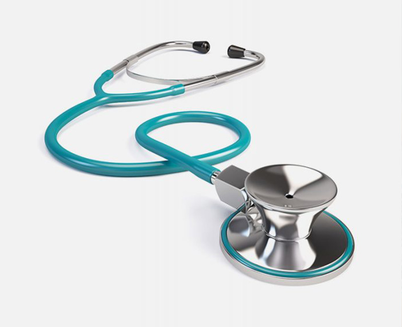
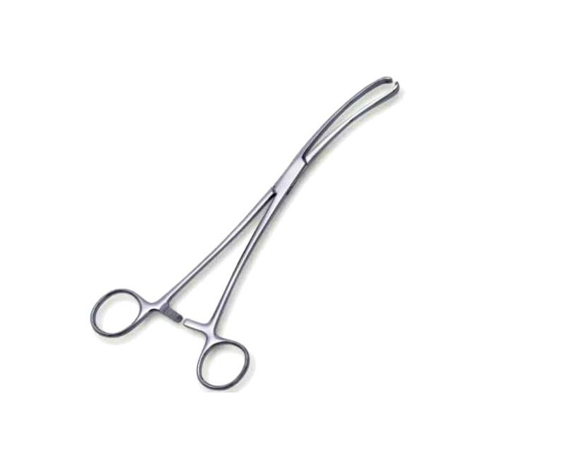
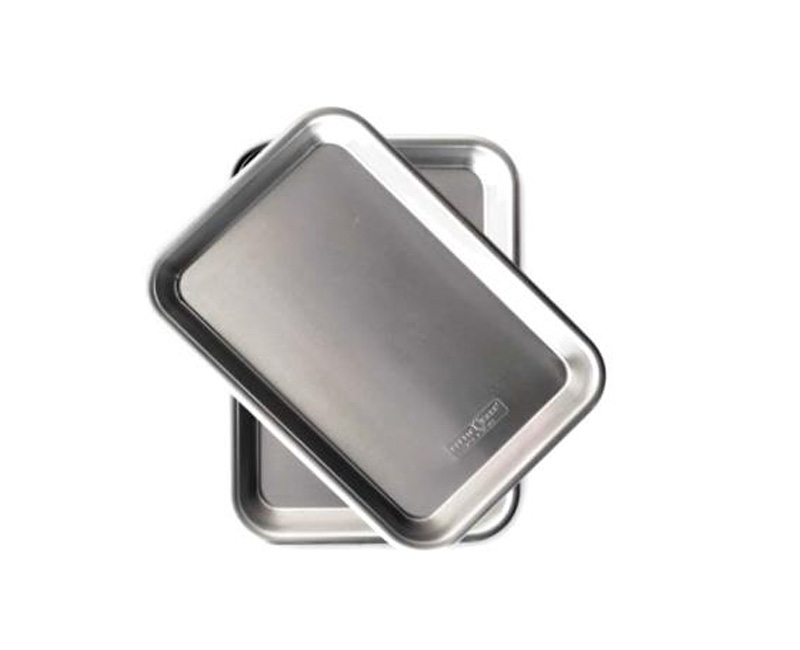
Visual reference cards from the source notes, including instrument images, names, and practical functions.
Fresh study guidance, clinical revision tips, and exam support written for nursing and midwifery students.
Move quickly from reading to practice with practical guides, diagnosis revision, and exam-ready downloads.
Focused pathways connect notes, blogs, quizzes, guides, and downloads around high-value nursing topics.
Choose a nursing or midwifery course and start learning.
🇺🇬 Proudly supporting nursing education across UgandaChoose from the four Nurses Online Discussion Notes courses. Each course opens into its curriculum, modules, topics, and available subtopics.
Complete curriculum outline and course list
Nursing medical instruments and equipment are essential tools that enable healthcare professionals to provide effective and efficient patient care.
Search all extracted Nurses Online Discussion Notes pages and open them inside the site subtopic structure.
Free downloads, nursing checklists, practical skills guides, exam preparation tools, and upcoming premium resources in one clean study hub.
Quick guides, checklists, and revision sheets for nursing and midwifery students.
Polished study packs, printable planners, case-study worksheets, and deeper clinical revision bundles are being prepared for review before release.
Subtopics, source coverage, and available images for this topic.

Free clinical notes, course outlines, quiz banks, and practical guides built for Uganda's nursing curriculum.

Continue learning, review quiz scores, and jump back into recent notes.
has completed the available Nurses Online Discussion learning notes and practice activities recorded in the learner dashboard.

Keep your course enrollment details current and review what the system is saving for your learning progress.
Registered learner accounts, course enrollment, and status.
Completed subtopics and course progress logs.
Total quiz attempts, average scores, and weak-area tracking.
Page visit logs plus Google Analytics & Search Console links.
Manage courses, modules, notes, resources, and instruments.
Review blog drafts, downloads, newsletter leads, quiz quality, and certificate readiness.
Create and edit section quizzes and subtopic quiz questions.
Upload and replace hero, course, subtopic, and instrument images.
Control quiz rails, subtopic banners, course ads, and mobile ad rules.
Logo paths, footer contact details, homepage media, and site copy.
Lock the public site during updates. Only /nod-admin and logged-in admins stay open.
Live management data appears here after admin login and database connection.
Choose a major nursing or midwifery section and practise with mixed ABCD questions, instant marking, and clear rationales.
Practice questions generated from the subtopic notes.
Patient care refers to the totality of care provided to individuals, families, or communities to attain, maintain or recover optimal health and quality of life. It is the core of nursing practice and involves the use of the nursing process in a compassionate and ethical manner.
The scope of patient care includes health promotion, prevention of illness, care of the sick, and rehabilitation. It encompasses physical, psychological, social, spiritual, and cultural dimensions of care.
Good patient care is safe, effective, respectful, timely and individualized.