Anatomy & Physiology
Subtopic:
The Lymphatic System
Learning Objectives
- Describe the anatomy and components of the lymphatic system, including lymph, lymphatic capillaries, vessels, trunks, and ducts.
- Differentiate between primary and secondary lymphatic organs, and explain their roles in immune function.
- Explain the functions and structural organization of the lymph nodes, including their layers, sinuses, and flow of lymph.
- Identify and describe the role of key organs in the lymphatic system: thymus, spleen, tonsils, and lymphatic nodules.
- Explain the structure and function of lymph, including its composition (interstitial fluid, WBCs, chyle) and directional flow.
- Understand the immune roles of lymphoid tissues, including their involvement in filtration, maturation, and response to pathogens.
- Describe major lymphatic trunks and ducts, and identify their drainage pathways into venous circulation.
- Recognize common lymphatic disorders and abnormalities, such as lymphadenitis, lymphangitis, elephantiasis, lymphomas, lymphedema, tonsillitis, and lymphosarcoma.
- The lymphatic system has some organs associated with it and these are divided into primary organs which are sites where stem cells divide and become immune-competent that is capable of mounting an immune response and these include: the thymus and red bone marrow and the secondary organs which are sites where maturation occurs and most immune responses occur. These include the lymph nodes, spleen, lymphatic nodules and the tonsils.
- It is through these organs that lymph flows. Lymph is a fluid formed when some components of blood plasma filter through blood capillary walls to form interstitial fluid. It mainly consists of interstitial fluid, white blood cells and chyle which is a lipoprotein complex absorbed into lymph from digested foods in the gut. Lymph flows in one direction and is a clear pale- yellow fluid but appears creamy white in the small intestines because of the fatty acids.
- Lymph vessels begin as lymphatic capillaries that unite to format larger lymphatic vessels that further unite to form trunks then ducts. Lymphatic capillaries are slightly larger than blood capillaries and have a special structure that permits interstitial fluid to flow into them but not out.
- This is so because the endothelial cells that make up the wall of the lymphatic capillary overlap in such a way that when pressure in the interstitial space is greater, the cells separate slightly to let in the interstitial fluid but when the pressure is greater in the lymphatic capillaries, the cells adhere more closely and thus lymph cannot escape back.
The organization of some the organs of lymphatic system are discussed below;
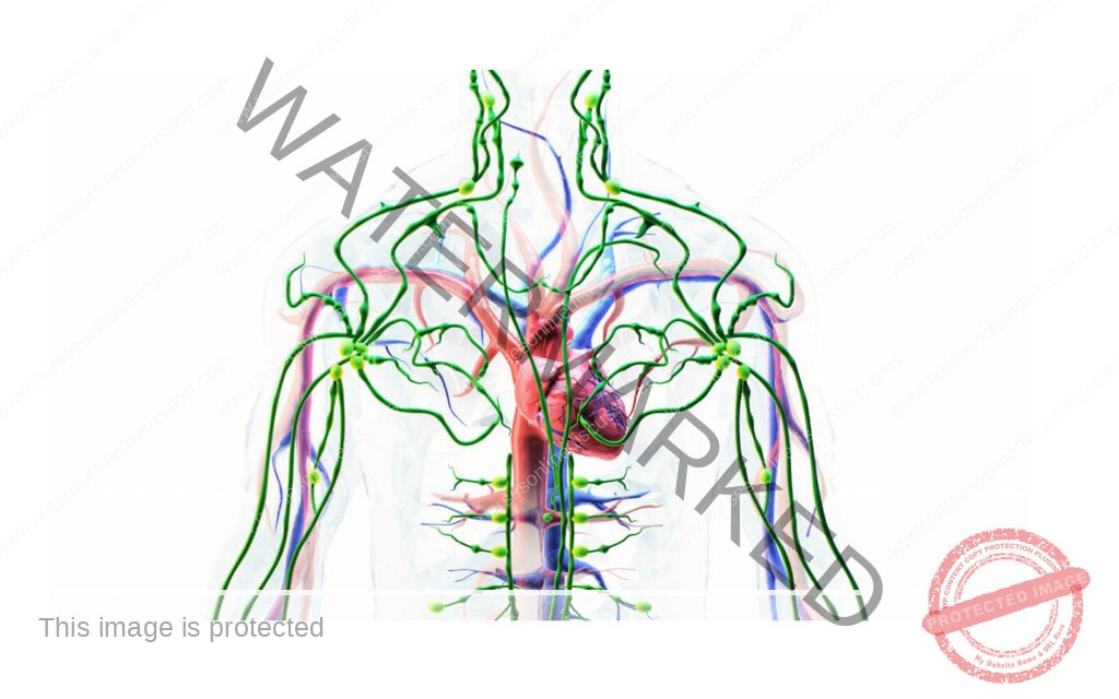
- Lymph nodes: these are located along the lymphatic vessels and are scattered throughout the body both superficially and deep. They usually occur in groups and large groups of these are specially found in mammary glands, axilla and groin.
- The lymph node has two sides that is the convex and the concave also known as hilum. The hilum is made up the vein, artery and efferent lymphatics and the convex is made of the afferent lymphatics. The lymph nodes have channels known as sinuses.
- The lymph node is organized in layers and these begin with the outermost capsule which radiates inward to form extensions known as trabeculae and the spaces between these trabeculae are known as the cortex. The central portion left is known as the medulla.
- The lymph thus travels through the lymph nodes from the afferent lymphatics then to the sub capsular sinuses to the trabecular sinuses that are adjacent to the trabeculae then onto the medullary sinuses the formed betwixt the channels left by the mesh like network by the medulla and from here the lymph is drained into the efferent vessel. The unidirectional movement here is maintained by the valves and the force to initiate the movement of the lymph across is from the reaction of the smooth muscles lining to arterial pulse and constant squeezing of skeletal muscles.
- Tonsils: these include adenoids, tubal, palatine and inguinal. Together these form a ring of lymphoid tissue around the throat. They mostly serve to trap pathogens from the food we eat and the air we inhale.
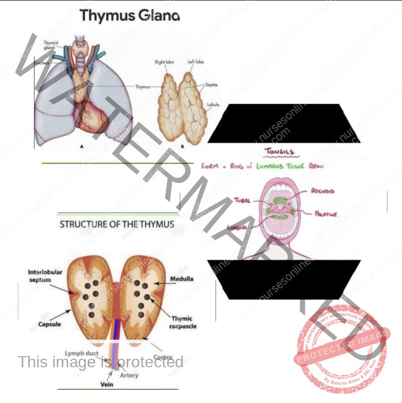
Thymus: this is a bi lobed organ found in the mediastinum behind the sternum and superior to the heart. It is the primary site for T – cell maturation and as such plays a major role in prevention of auto immunity by destroying T – cells that react to self antigens. The thymus as an enveloping layer of connective tissue that holds the two lobes closely together but a connective capsule encloses each lobe separately. Extensions of the capsule known as trabeculae divide each lobe into lobular. The thymus is most active in infants and is also a reservoir in that it keeps erythrocytes and platelets incase of any emergencies.
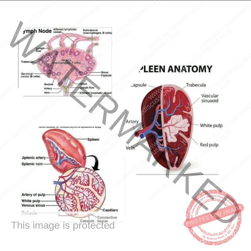
Spleen: this is the largest mass of lymphatic tissue in the body and is located in the left hypochondriac region below the diaphragm and above the stomach. The spleen has red pulp and white pulp. Red pulp acts as a filtration unit where old and damaged erythrocytes and bacteria are destroyed. White pulp is a site where all lymphoid cells reside and interact un the skin. It’s also where antibody coated bacteria are filtered out and antibodies made by B – cells.
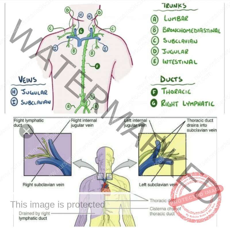
There are trunks into which lymph is drained into and these are 2 lumbar trunks, 2 broncho-mediatinal trunks, 2 subclavian trunks, 2 jugular trunks and 1 intestinal trunk. For here lymph is delivered into ducts that is the right lymphatic duct that drains the right arm and right side of the head and chest and the thoracic duct that collects lymph from the rest of the body. From here the right lymphatic duct dumps blood at the junction of the right subclavian vein and right jugular vein while the thoracic duct dumps at the same Junction the left side of the body. This is so because at this spot there’s less pressure so it’s convenient.
The common abnormalities of the lymphatic system
- The lymphatic system is involved with the production of lymphocytes which fight infectious diseases and lymphatic system filters blood and lymph remove microorganisms. Lymphopathy is the disorder of the lymphatic system
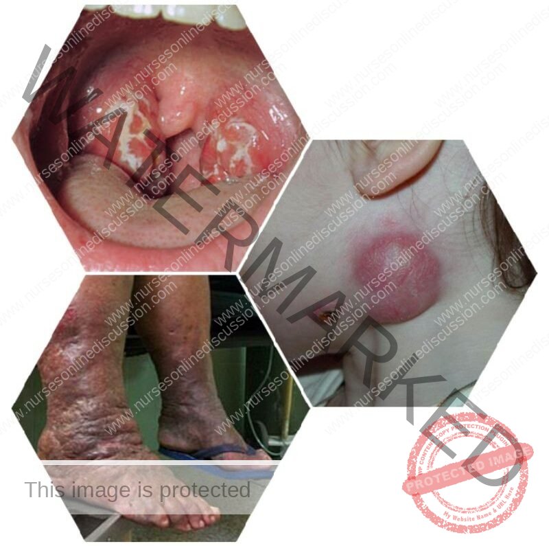
- Lymphadentis: ls an inflammation of the lymphatic nodes which causes them to become enlarged and tender. This inflammation is an indication that microorganisms are being trapped and destroyed within the lump nodes
- Lymphangetis: this results in visible red streaks in the skin that extend away from the site of infection
- Elephantiasis: ls caused by long, slender roundworms (U here ria bancrofti) The adult worms lodge in the lymphatic vessels and block lymph flow.The resulting accumulation of fluid in the interstitial space and lymphatic vessels can cause permanent swelling and enlargement of the limb.The offspring of the adult worms pass through the lymphatic system into the blood from which they can be transferred to anothehuman by mosquitoes
- Lymphoma is a neoplasm (tumor) of lymphatic tissue. Lymphomas are divided into two; Hodgkin disease and Non Hodgkin Lymphomas
- Lymphomas begin as an enlarged painless mass of lymph nodes, they depress the immune system and the patient is susceptible to infections. Enlargement of the lymph nodes also compress surrounding structures and produce complications
- Lymphedema: Swelling due to accumulation of lymphatic fluid in the interstitial space
- Tonsillitis: congestion of the tonsils typically with bacteria which causes them to become red, swollen and sore
- Lymphosarcoma; cancer within the lymphatic tissue
Summary
The lymphatic system communicates with the blood circulatory system and is closely associated with it. It consists of:
- Lymphatic capillaries made of endothelium (simple squamous epithelium)
- Lymphatic vessels made of three layers like veins; also they have valves.
- Lymphatic ducts: are ducts that drain different parts of the body and includes:
- Right lymphatic duct drains upper right part of the body and empties in to right subclavian vein
- Thoracic duct drains remainder part of the body and empties into left subclavian vein.
- Lymph is the fluid within the lymphatic capillaries and vessels; which is derived from tissue fluid. Tissue fluid is derived from the blood plasma. A certain amount of this fluid and waste products from the cells is returned to the venous capillaries, but within the tissue spaces fine capillary vessels known as lymphatic capillaries begin, which help to drain the waste products and water from the interstitial spaces. Also larger sized materials or substances of the result of phagocytosis of pathogenic micro-organisms are drained away in the lymphatic capillaries and vessels.
- Lymphoid tissues are distributed throughout the body.
These are:
- Lymph nodes help for filtration of lymph
- Tonsils also help for filtration of tissue fluids
- Thymus for processing of T-lymphocytes and hormone(thymosin) secretion to stimulate T-lymphoid tissues.
- Spleen helps for filtration of blood, and destruction of old red cells.
Join Our WhatsApp Groups!
Are you a nursing or midwifery student looking for a space to connect, ask questions, share notes, and learn from peers?
Join our WhatsApp discussion groups today!
Join NowWe are a supportive platform dedicated to empowering student nurses and midwives through quality educational resources, career guidance, and a vibrant community. Join us to connect, learn, and grow in your healthcare journey
Quick Links
Our Courses
Legal / Policies
Get in Touch
(+256) 790 036 252
(+256) 748 324 644
Info@nursesonlinediscussion.com
Kampala ,Uganda
© 2026 Nurses online discussion. All Rights Reserved