Anatomy & Physiology I
Subtopic:
The Digestive System
Learning Objectives
- Define digestion and absorption, and describe their significance in nutrient processing.
- Identify components of the digestive tract and accessory organs.
- Describe the structure and function of key digestive organs: mouth, pharynx, esophagus, stomach, small intestine, and large intestine.
- Explain the layers of the digestive tract and their role in peristalsis.
- Understand roles of salivary glands, liver, gallbladder, and pancreas in digestion.
- Describe chemical digestion of carbohydrates, proteins, and fats, and match enzymes with their substrates.
- Explain nutrient absorption mechanisms via villi/microvilli and water/osmotic transport.
- Recognize common digestive disorders and the impact of antibiotics on gut health.
General Function
Everybody cell needs a constant supply of nutrients to provide energy and building blocks for the manufacture of body substances. Food as we take it in, however, is too large to enter the cells. It must first be broken down into particles small enough to pass through the cell membrane. This process is known as digestion. After digestion, food must be carried to the cells in every part of the body by the circulation.
The transfer of food into the circulation is called absorption.
Digestion and absorption are the two chief functions of the digestive system.
Structure and Function of Organs of Digestion and Accessory Organs
For our purpose the digestive system may be divided into two groups of organs:
- The digestive tract, a continuous passageway beginning at the mouth, where food is taken in, and terminating at the anus, where the solid waste products of digestion are expelled from the body
- The accessory organ, which are necessary for the digestive process but are not a direct part of the digestive tract. They release substances into the digestive tract through ducts.
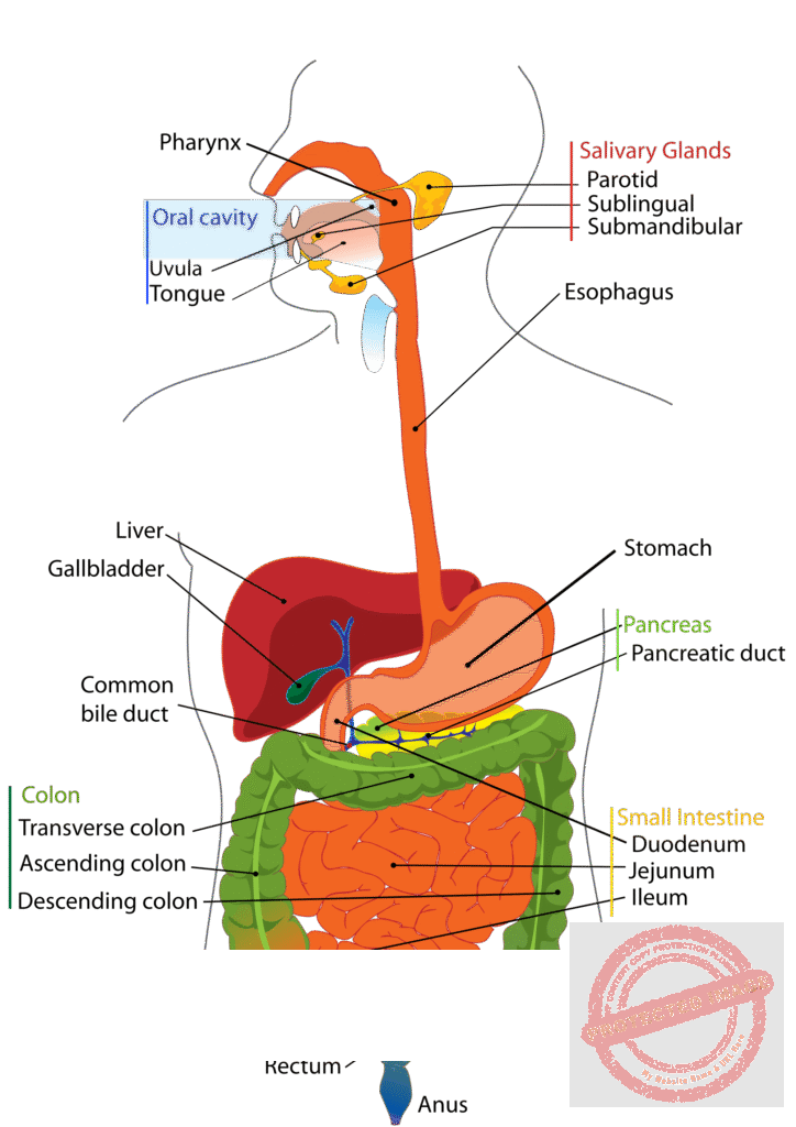
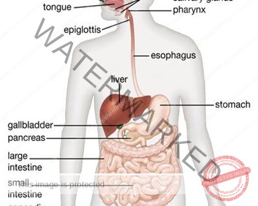
The Digestive Tract:
- The digestive tract is a muscular tube extending through the body. It is composed of several parts: the mouth, pharynx, esophagus, stomach, small intestine, and large intestine. The digestive tract is sometimes called the alimentary tract, derived from a Latin word that means “food”.
- It is more commonly referred to as the gastrointestinal (Gl) tract because of the major importance of the stomach and intestine in the process of digestion.
The Walls of the Digestive Tract
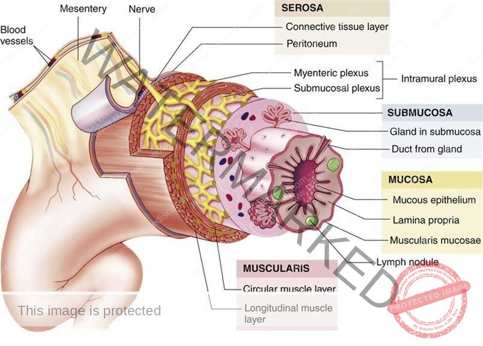
Although specified for specific tasks in different organs, the wall of the digestive tract, from the esophagus to the anus, is similar in structure throughout. Follow the diagram of the small intestine in from the innermost to the outermost surface.
First is the mucous membrane, so called because its epithelial layer contains many mucus-secreting cells. The type of epithelium is simple columnar.
The layer of connective tissue beneath this, the submucosa, contains blood vessels and some of the nerves that help regulate digestive activity. Next are two layers of smooth muscle. The inner layer has circular fibers, and the outer layer has longitudinal fibers.
The alternate contractions of these muscles create the wavelike movement that propels food through the digestive tract and mixes it with digestive juices. This movement is called peristalsis. The outermost layer of the wall consists of fibrous connective tissue. Most of the abdominal organs have an additional layer of serous membrane that is part of the peritoneum.
The Peritoneum
The abdominal cavity is lined with a thin, shiny serous membrane that also covers most of the abdominal organs. The portion of this membrane that lines the abdomen is called the parietal peritoneum; that covering the organ is called the visceral peritoneum.
In addition to these single layered portions of the peritoneum there are a number of double-layered structures that carry blood vessels, lymph vessels, and nerves, and sometimes act as ligaments supporting the organs. The mesentery is a double-layered portion of the peritoneum shaped somewhat like a fan.
The handle portion is attached to the back wan, and the expanded long edge is attached to the small intestine. Between the two layers of membrane that fOl1ll the mesentery are the blood vessels, lymphatic vessels, and nerves that supply the intestine. The section of the peritoneum that extends from the colon to the back wall is the mesocolon.
A large double layer of the peritoneum containing much fat hangs like an apron over the front of the intestine. This greater omentum extends from the lower border of the stomach into the pelvic part of the abdomen and then loops back up to the transverse colon. There is also a smaller membrane, called the lesser omentum that extends between the stomach and the liver.
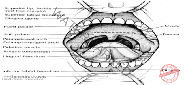
The Mouth
The mouth, also called the oral cavity, is where a substance begins its travels through the digestive tract. The mouth has three digestive functions:
- To receive food, a process called ingestion
- To prepare food for digestion
- To begin the digestion of starch.
In to this space projects a muscular organ, the tongue, which is used for chewing and swallowing, and is one of the principal organs of speech. The tongue has on its surface a number of special organs, called taste buds, by means of which taste sensations (bitter, sweet, sour, or salty) can be differentiated.
The oral cavity also contains the teeth. A child between 2 and 6 years of age has 20 teeth; an adult with a complete set of teeth has 32. Among these, the cutting teeth, or incisors, occupy the front part of the oral cavity, whereas the lager grinding teeth, the molars, are in the back.
The mouth, showing the teeth and tonsils (see the diagrams in your text book)
Deciduous or Baby Teeth:
The first eight deciduous teeth to appear through the gums are the incisors. Later the canines (eye teeth) and molar appear. Usually, the 20 baby teeth have all appeared by the time a child has reached the age of 2 or 21/2 years. During the first 2 years the permanent teeth develop within the jawbones from buds that are present at birth. The first permanent tooth to appear is the important 6-year molar. This permanent tooth comes in before the baby incisors are lost. Because decay and infection of adjacent deciduous molars may spread to and involve new, permanent teeth, deciduous teeth need proper care.
Permanent Teeth
As a child grows, the jawbones grow, making space for additional teeth. After the 6-year molars have appeared, the baby incisors loosen and are replaced by permanent incisors.
Next, the baby canines (cuspids) are replaced by permanent canines, and finally, the baby molars are replaced by the bicuspids (premolars) of the permanent teeth.
Now the larger jawbones are ready for the appearance of the 12-year, or second, permanent molar teeth. During or after the late teens, the third molars, or so-called wisdom teeth, may appear. In some cases, the jaw is not large enough for these teeth or there are other abnormalities, so that the third molars may have to be removed.
The Salivary Glands
While food in the mouth, it is mixed with saliva, one purpose of which is to moisten the food and facilitate the processes of chewing, or mastication, and swallowing, or deglutition.
Saliva also helps keep the teeth and mouth clean and reduce bacterial growth.
This watery mixture contains mucus and an enzyme called salivary amylase, which begins the digestive process by converting starch to sugar. It is manufactured mainly by three pairs of glands that function as accessory organs:
- The parotid glands, the largest of the group, are located below and in front of the ear.
- The submandibular, or submaxillary, glands are located near the body of the lower jaw
- The sublingual glands are under the tongue.
- All these glands empty by means of ducts into the oral cavity.
The Pharynx and Esophagus:
- The pharynx is commonly referred to as the throat. The oral part of the pharynx is visible when you look into an open mouth and depress the tongue. The palatine tonsils may be seen at either side. The pharynx also extends upward to the nasal cavity and downward to the level of the larynx. The soft palate is tissue that forms the back of the roof of the oral cavity. From it hangs a soft, fleshy, V -shaped mass called the uvula.
- In swallowing, a small portion of chewed food mixed with saliva, called a bolus, is pushed by the tongue into the pharynx, swallowing occurs rapidly by an involuntary reflex action. At the same time, the soft palate and uvula are raised to prevent food and liquid from entering the nasal cavity, and the tongue is raised to seal the back of the oral cavity.
- The entrance of the trachea is guarded during swallowing by a leaf-shaped cartilage, the epiglottis, which covers the opening of the larynx. The swallowed food is then moved by peristalsis into esophagus, a muscular tube about 25cm (10 inches) long that carries food into the stomach. No additional digestion occurs in the esophagus.
- Before joining the stomach, the esophagus must pass through the diaphragm. It passes through a space in the diaphragm called the esophageal hiatus. If there is a weakness in the diaphragm at this point, a portion of the stomach or other abdominal organ may protrude through the space, a condition called hiatal hernia.
The Stomach
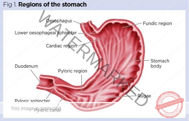
The stomach is an expanded J-shaped organ in the upper left region of the abdominal cavity
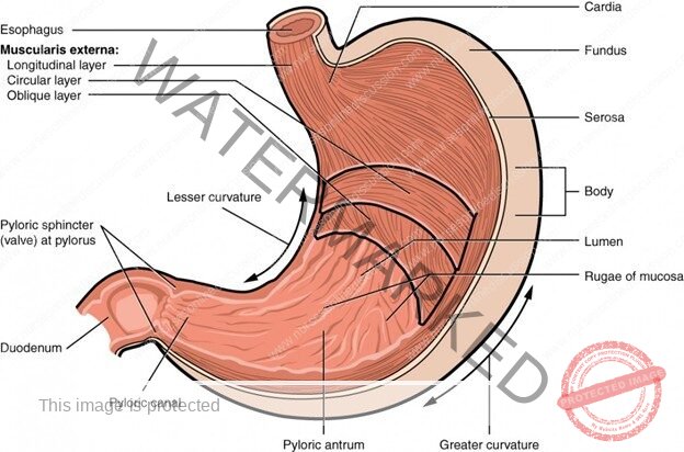
- In addition to the two muscle layers already described, it has a third, inner oblique (angled) layer that aids in grinding food and mixing it with digestive juices. The left-facing arch of the stomach is the greater curvature, whereas the right surface forms the lesser curvature. Each end of the stomach is guarded by a muscular ring, or sphincter, that permits the passage of substances in only one direction.
- Between the esophagus and the stomach is the lower esophageal sphincter (LES). This valve has also been called cardiac sphincter because it separates the esophagus from the region of the stomach that is close to the heart.
- We are sometimes aware of the existence of this sphincter; sometimes it does not relax as it should, produce a feeling of being unable to swallow past that point. Between the distal, or far, end of the stomach and the small intestine is the pyloric sphincter. The region of the stomach leading into this sphincter, the pylorus, is important in regulating how rapidly food moves into the small intestine.
The stomach serves as a storage pouch, digestive organ, and churn. When the stomach is empty, the lining forms many folds called rugae. These folds disappear as the stomach expands. (It may be stretched to hold one half of a gallon of food and liquid.) Special cells in the lining of the stomach secrete substances that mix together to form gastric juice, the two main components of which are:
- Hydrochloric acid (HCL), a strong acid that softens the connective tissue in meat and destroys foreign organisms
- Pepsin, a protein-digesting enzyme. This enzyme is produced in an inactive form and is activated only when food enters the stomach and HCL is produced.
The semi-liquid mixture of gastric juice and food that leaves the stomach to enter the small intestine is called chyme.
The Small Intestine
- The small intestine is the longest part of the digestive tract. It is known as the small intestine because, although it is longer than the large intestine, it is smaller in diameter, with an average width of about 2.5 cm (1 inch). When relaxed to its full length, the small intestine is about 6 m (20 feet) long. The first 25 cm (10 inches) or so of the small intestine make up the duodenum. Beyond the duodenum are two more divisions: the jejunum, which forms the next two fifths of the small intestine, and the ileum, which constitutes the remaining portion.
- The wall of the duodenum contains glands that secrete large amounts or mucus to protect the small intestine from the strongly acid chyme entering from the stomach. Cells of the small intestine also secrete enzymes that digest proteins and carbohydrates. In addition, digestive juices from the liver and pancreas enter the small intestine through a small opening in the duodenum. Most of the digestive process takes place in the small intestine under the effects of these juices.
- Most absorption of digested food also occurs through the walls of the small intestine. To increase the surface area of the organ for this purpose, the mucosa is formed into millions of tiny, finger-like projections, called villi, which give the inner surface a velvety appearance. In addition, each epithelial cell has small projecting folds of the cell membrane known as microvilli.
- These create a remarkable increase in the total surface area available in the small intestine for the absorption of nutrients.
The Large Intestines
- Any material that cannot be digested as it passes through the digestive tract must be eliminated from the body. In addition, most of the water secreted into the digestive tract for proper digestion must be reabsorbed into the body to prevent dehydration. The storage and elimination of undigested waste and the reabsorption of water are the functions of the large intestine.
- The large intestine is about 6.5 cm (2.5 inches) in diameter and about 1.5 m (5 feet) long. The outer longitudinal muscle fibers form three separate bands on the surface. These bands draw up the wall of the organ to give it its distinctive puckered appearance.
- The large intestine begins in the lower right region of the abdomen. The first part is a small pouch called the cecum.
- Between the ileum of the small intestine and the ceacum is a sphincter, the ileocecal valve that prevents food from traveling backward into the small intestine. Attached to the +m is a small, blind tube containing lymphoid tissue; it is called the vernriform appendix (vermiform means “wotmlike”).
- Inflammation of this tissue as a result of infection or obstruction is appendicitis.
- The second portion, the ascending colon, extends upward along the right side of the abdomen toward the liver. The large intestine then bends across the abdomen, forming the transverse colon. At this point it bends sharply and extends downward on the left side of the abdomen into the pelvis, forming the descending colon. The lower part of the colon bends posteriorly in an S shape and continues downward as the sigmoid colon. The sigmoid colon empties into the rectum, which serves as a temporary storage area for indigestible or unabsorbable food residue.
- Enlargement of the veins in this area constitutes haemorrhoids. A narrow portion of the distal large intestine is called the anal canal. This leads to the outside of the body through an opening called the anus.
- Large quantities of mucus, but no enzymes, are secreted by the large intestine. At intervals, usually after meals, the involuntary muscles within the walls of the large intestine propel solid waste material, called feces or stool, toward the rectum. This material is then eliminated from the body by both voluntary and involuntary muscle actions, a process called defecation.
- While the food residue is stored in the large intestine, bacteria that normally live in the colon act on it to produce vitamin K and some of the B-complex vitamins. As mentioned, systemic antibiotic therapy may destroy these bacteria and others living in the large intestine, causing undesirable side effects.
The Accessory Structures
The Liver (draw the structure)
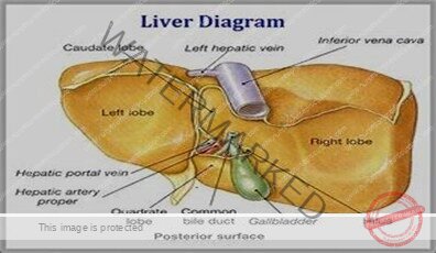
The liver, often referred to by the word root hepat, is the largest glandular organ of the body. It is located in the upper right portion of the abdominal cavity under the dome of the diaphragm. The lower edge of a normal-sized liver is level with the lower margin of the ribs.
The human liver is the same reddish brown color as the animal liver seen in the supeln1arket. It has a large right lobe and a smaller left lobe; the right lobe includes two inferior smaller lobes. The liver is supplied with blood through two vessels: the portal vein and the hepatic artery. These vessels deliver about 11/2 quarts of blood to the liver every minute. The hepatic artery carries oxygenated blood, whereas the portal system of veins carries blood that is rich in the end products of digestion. This most remarkable organ has so many functions that only some of its major activities can list here:
- The storage of glucose (simple sugar) in the form of glycogen, in animal starch. When the blood sugar level falls below normal, liver cells convert glycogen to glucose and release it into the bloodstream; this serves to restore the normal concentration of blood sugar.
- The formation of blood plasma proteins, such as albumin, globulins, and clotting factors
- The synthesis of urea, a waste product of protein metabolism. Urea is released into the blood and transported to the kidneys for elimination.
- The modification of fats, so cells all over the body can use them more efficiently
- The manufacture of bile
- The destruction of old red blood cells. The pigment released from these cells in both the liver and the spleen is eliminated in the bile. This pigment (bilirubin) gives the stool its characteristic dark color.
- The detoxification (removal of the poisonous properties) of harmful substances such as alcohol and certain drugs
- The storage of some vitamins and iron
- The main digestive function of the liver is the production of bile. The salts contained in bile act like a detergent to emulsify fat, that is, to break up fat into small droplets that can be acted on more effectively by digestive enzymes. Bile also aids in the absorption of fat from the small intestine. Bile leaves the lobes of the liver by two ducts that merge to foln1 the common hepatic duct. After collecting bile from the gallbladder, this duct, now called common bile duct, delivers bile into the duodenum.

The Gallbladder
The gallbladder is a muscular sac on the inferior surface of the liver that serves as a storage pouch for bile. Although the liver may manufacture bile continuously, the body is likely to need it only a few times a day. Consequently, bile from the liver flows into the hepatic ducts and then up through the cystic duct connected with the gallbladder. When chyme enters the duodenum, the gallbladder contracts, squeezing bile through the cystic duct and into the common bile duct leading to the duodenum.
The Pancreas
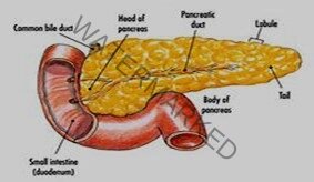
- The pancreas is a long gland that extends from the duodenum to the spleen. The pancreas produces enzymes that digest fats, proteins, carbohydrates, and nucleic acids. The protein digesting enzymes are produced in inactive forms, which must be converted to active forms in the small intestine by other enzymes. The pancreas also produces large amounts of alkaline fluid, which neutralizes the chyme in the small intestine, thus protecting the lining of the digestive tract.
- These juices collect in a main duct that joins the common bile duct or empties into the duodenum near the common bile duct. Most persons also have an additional smaller duct that opens into the duodenum. Because they are usually confined to proper channels, pancreatic enzymes do not damage body tissues.
- However, if the bile ducts become blocked, pancreatic enzymes back up into the pancreas. Also, in some cases of gallbladder disease, disease, infection may extend to the pancreas and cause abnormal activation of the pancreatic enzymes. In either circumstance, the pancreas suffers destruction by its own juice, and the outcome can be fatal; this condition is known as acute pancreatitis.
- The pancreas also functions as an endocrine gland, producing the hormones insulin and glucagon that regulate sugar metabolism. These secretions of the islet’s cells are released directly into the blood.
Digestion and Absorption of Carbohydrates, Fats, and Proteins
Digestion
- Digestion, a complex process that occurs in the alimentary canal, consists of physical and chemical changes that prepare food for absorption. Mechanical digestion breaks food into tiny particles, mixes them with digestive juices, moves them along the alimentary canal, and finally eliminates the digestive wastes from the body.
- Chewing or mastication, swallowing or deglutition, peristalsis, and defecation are the main processes of mechanical digestion. Chemical digestion breaks down large, non-absorbable food molecules−molecules that are able to pass through the intestinal mucosa into blood and lymph.
- Chemical digestion consists of numerous chemical reactions catalyzed by enzymes in saliva, gastric juice, pancreatic juice, and intestinal juice.
Carbohydrate Digestion
- Very little digestion of carbohydrates (starches and sugars) occurs before food reaches the small intestine. Salivary amylase usually has little time to do its work because so many of us swallow our food so fast. Gastric juice contains no carbohydrate-digesting enzymes. But after the food reaches the small intestine, pancreatic and intestinal juice enzymes digest the starches and sugars. A pancreatic enzyme (amylase) starts the process by changing starches into a double sugar, namely, maltose. Three intestinal
- enzymes−rnaltase, sucrase, and lactase−digest double sugars by changing them into simple sugars, chiefly glucose (dextrose). Maltase digests maltose (malt sugar), sucrose digests sucrose (ordinary cane sugar), and lactase digests lactose (milk sugar). The end product of carbohydrate digestion is the so-called simple sugar; the most abundant is glucose.
Protein Digestion
- Protein digestion starts in the stomach. Two enzymes (rennin and pepsin) in the gastric juice cause the giant protein molecules to break up into somewhat simpler compounds. Pepsinogen, a component of gastric juice, is converted into active pepsin enzyme by hydrochloric acid (also in gastric juice). In the intestine, other enzymes (trypsin in the pancreatic juice and peptidases in the intestinal juice) finish the job of protein digestion.
- Every protein molecule is made up of many amino acids joined together. When enzymes have split up the large protein molecule into its separate amino acids, protein digestion is completed. Hence the end product of protein digestion is amino acids. For obvious reasons, the amino acids are also referred to as protein building blocks.
Fat Digestion
Very little carbohydrate and fat digestion occurs before food reaches the small intestine. Most fats are undigested until after emulsification by bile in the duodenum (that is, fat droplets are broken into very small droplets). After this takes place, pancreatic lipase splits up the fat molecules into fatty acids and glycerol (glycerine). The end products of fat digestion, then, are fatty acids and glycerol.
the main facts about chemical digestion:
Enzyme names indicate the type of food digested by the enzyme. For example, the name amylase indicates that the enzyme digests carbohydrates (starches and sugars), protease indicates a protein- digesting enzyme, and lipase means a fat-digesting enzyme. When carbohydrate digestion has been completed, starches (polysaccharides) and double sugars (disaccharides) have been changed mainly to glucose, a simple sugar (monosaccharide).
The end products of protein digestion, on the other hand, are amino acids. Fatty acids and glycerol are the end products of fat digestion.
Absorption
After food is digested, it is absorbed; that is, it moves through the mucous membrane lining of the small intestine into the blood and lymph. In other words, food absorption is the process by which molecules of amino acids, glucose, fatty acids, and glycerol goes from the inside of the intestines into the circulating fluids of the body. Absorption of foods is just as essential as digestion of foods. The reason is fairly obvious. As long as food stays in the intestines, it cannot nourish the millions of cells that compose all other parts of the body. Their lives depend on the absorption of digested food and its transportation to them by the circulating blood.
Structural adaptations of the digestive tube, including;
- Folds in the lining mucosa, villi, and micro villi, increase the absorptive surface and the efficiency and speed of transfer of materials from the intestinal lumen to body fluids.
- Many salts such as sodium are actively transported through the intestinal mucosa.
- Water follows by osmosis. Other nutrients are also actively transported into the blood of capillaries in the intestinal villi.
- Fats enter the lymphatic vessels or lacteals found in intestinal villi.
Assignment : Give a summery of Chemical digestion
Join Our WhatsApp Groups!
Are you a nursing or midwifery student looking for a space to connect, ask questions, share notes, and learn from peers?
Join our WhatsApp discussion groups today!
Join NowWe are a supportive platform dedicated to empowering student nurses and midwives through quality educational resources, career guidance, and a vibrant community. Join us to connect, learn, and grow in your healthcare journey
Quick Links
Our Courses
Legal / Policies
Get in Touch
(+256) 790 036 252
(+256) 748 324 644
Info@nursesonlinediscussion.com
Kampala ,Uganda
© 2026 Nurses online discussion. All Rights Reserved